How Long It Takes a Lab to Analyze Mole Biopsy for Melanoma
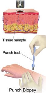
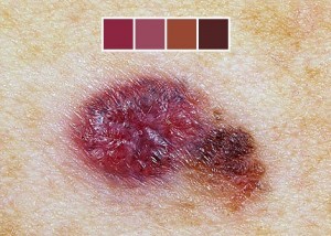
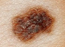
When a dermapathologist views mole cells under a microscope, how long does it take them to identify if it’s melanoma or benign?
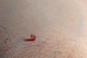
Your doctor will tell you it takes a week to two weeks for “the results to come back.” Sometimes the doctor will say it’ll take “a couple days.” (more…)
Can a Mole Be Too Tiny to Biopsy for Melanoma?
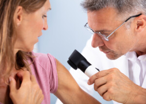
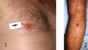
If your dermatologist told you that a mole is too tiny to be biopsied, and to wait a little longer to see if it gets bigger, get a second opinion. (more…)
Is the 16:8 Diet Just Another Passing Fad?
Let’s get the straight & narrow on the 16:8 diet: Is it effective or just another hyped up fad?
In the 16:8 diet, you eat whatever you want for eight hours only during a given day, and then absolutely nothing (other than water and zero-calorie drinks) for the other 16 hours. No exceptions.
The hours of feasting are from 10 am to 6 pm.
The hours of fasting are from 6:01 pm to 5:59 am.
Sounds easy enough, right?
Well, according to a study of 23 obese people (average age 45, average body mass index 35), the 16:8 diet has merit.
The participants followed the 16:8 diet for 12 weeks and lost weight plus improved their blood pressure.
• On average they ate 350 fewer calories/day.
• The average weight loss was three percent of their body weight.
• This is the first study of the 16:8 diet, and the full report is in Nutrition and Healthy Aging (June 2018).
During the eight hours that one can eat, there are no limits on type of food or quantity.

Shutterstock/Hurst Photo
On one hand, it’s easy to see why there was some weight loss. Many overeaters do much of their feasting after they get home from work – the very hours that are off-limits from eating on the 16:8 diet.
But on the other hand, knowing that you can’t eat even a bite between 6 pm and bedtime might encourage more overeating earlier in the day to avoid feeling hungry later in the evening.
Overall, though, for the 23 participants, this did not happen enough to thwart a collective weight loss over 12 weeks.
Another point to consider is that for some individuals, most overeating occurs at home for several reasons, and most people are away from home, at work between 10 am and 6 pm — where binge eating can be impossible with certain jobs.
Being at home, watching evening TV shows, means a strong conditioned stimulus for overeating.
The study authors do not recommend that the 16:8 diet be a permanent solution, as it still encourages overeating, and includes unlimited amounts of junk food.
Sustainability of the 16:8 Diet
“16:8 is one of the many versions of intermittent fasting which is not sustainable in the long run,” says Shana Spence, MS, RDN, CDN, a registered dietitian nutritionist based in New York, and was not involved with the study.
“There are some researched benefits to fasting — weight loss being one. Yes, you’ll definitely lose weight if you’re restricting your intake — but is it sustainable? I don’t think so.
“As for my overall opinion, the idea of limiting yourself to an eight hour window seems like an extreme.”
 Shana Spence of The Nutrition Tea is committed to providing trending information and nutrition facts covering a wide range including nutrition for heart disease and diabetes, pediatric nutrition and healthful lifestyles.
 Lorra Garrick has been covering medical, fitness and cybersecurity topics for many years, having written thousands of articles for print magazines and websites, including as a ghostwriter. She’s also a former ACE-certified personal trainer.
.
Top image: ©Lorra Garrick
Source: sciencedaily.com/releases/2018/06/180618113038.htm
Why Your Child’s Poop Smells Like Mothballs

You don’t even have mothballs in the house, so what makes your child’s poop stink like mothballs?
“Foul smelling stool is a byproduct of gut inflammation and dysbiosis [a state of imbalance in the gut’s natural bacterial population],” says Joel Gator Warsh, MD, of Integrative Pediatrics and Medicine, Studio City, CA, and part of the pediatric staff of Cedars-Sinai Hospital.
“If your child’s poop smells like mothballs, I would consider a food sensitivity or allergy.
“The inflammation in the gut and the undigested food particles can make your child’s bowel movement be very foul smelling.
“One description of this smell is a musty ‘mothball’ odor. The most common allergies leading to this smell are gluten/wheat and dairy, so I would consider removing those from the diet and see if the smell changes.”
This means if a loaf of gluten-containing bread was on a cutting board, do NOT use that cutting board to cut up something that’s gluten-free such as fresh vegetables.
Even one crumb of gluten, in someone with a gluten sensitivity, can mess things up.
“You can also be lactose intolerant and that causes abdominal pain, bloating, severe flatulence and foul smelling, mothball stools,” says Dr. Warsh.
Give your child lactose-free milk (if they already drink milk) and see what happens.
“I would also consider having stool tested for bacterial, viral and fungal pathogens.
“An infection in the gut can lead to changes in stool consistency, smell and size.
“Infections with parasites can be particularly worrisome and can create distinctive, odd odors.”
In the U.S., parasite infections in kids are relatively uncommon — but still possible.
The risk heightens in areas with poor sanitation or close contact with contaminated soil or untreated water.
Pinworms are the most frequent, while hookworms and giardia occur less often.
Good hygiene and proper handwashing significantly lower risk.
Nevertheless, a parasitic infection causing a mothball odor to BMs ranks very low on the list of possible explanations.
- The parent’s subjective interpretation of the odor can resemble that of mothballs.
 Dr. Warsh and his Studio City, Los Angeles clinic treat a wide array of common pediatric issues using holistic and conventional treatments. He works with nutritionists, naturopaths, Ayurvedic practitioners, acupuncturists and more.
 Lorra Garrick has been covering medical, fitness and cybersecurity topics for many years, having written thousands of articles for print magazines and websites, including as a ghostwriter. She’s also a former ACE-certified personal trainer.
.
Top image: Shutterstock/Anatoliy Karlyuk
What Causes a Baby to Drag a Leg when Crawling?

A baby may have a dragging leg from the time they begin crawling, or, it may develop at some point after crawling has become habitual.
“Motor developmental milestones vary greatly from child to child,” begins Joel Gator Warsh, MD, of Integrative Pediatrics and Medicine, Studio City, CA, and part of the pediatric staff of Cedars-Sinai Hospital.
“No two are exactly the same. Some children crawl at seven months, others never crawl.”
That second group of babies amazingly go from not crawling to actual pitter-pattering.
When a Crawling Baby Drags a Foot…
Dr. Warsh continues, “Favoring one leg when crawling is usually normal. Babies do what works, so some scoot, and some use just one side, while others master the bilateral skill more quickly.
“If you notice a baby favoring one side or not using one limb, it is important to have a thorough neurologic exam at your doctor’s to insure that the signals from the brain are getting sent appropriately to the leg.
“Nerves, tendons and muscles can all be injured or damaged, leading a child to favor one side.
“Most of these issues will be noticed after birth, but it is possible that they can develop at a later age or not be noticed until motor skills are tested at an older age.
“Another common injury is a toddler fracture. If you suddenly notice a child not using one limb, consider a fracture.”
Do not try to diagnose the absence of a fracture, and do not move the affected limb in an attempt to see if it brings out pain. Bring the baby to a doctor.
“Babies and toddlers can get injured, even when we are not looking, so just because there wasn’t a known trauma, does not mean a fracture couldn’t occur. Obtain an X-ray to rule out an occult fracture.”
And it needs to be pointed out that a fracture could have been caused by a caretaker (teen babysitter, nanny, even older sibling) of the baby.
 Dr. Warsh and his Studio City, Los Angeles clinic treat a wide array of common pediatric issues using holistic and conventional treatments. He works with nutritionists, naturopaths, Ayurvedic practitioners, acupuncturists and more.
 Lorra Garrick has been covering medical, fitness and cybersecurity topics for many years, having written thousands of articles for print magazines and websites, including as a ghostwriter. She’s also a former ACE-certified personal trainer.
.
Top image: Freepik.com, freepic.diller
Can a Preschooler Deliberately Withhold Poops for 3 Weeks?
Is it really possible for a preschool child to deliberately hold in his or her bowel movements for up to three weeks?
“If a preschooler truly has not had a BM for three weeks, I would be concerned and seek medical care immediately,” says Joel Gator Warsh, MD, of Integrative Pediatrics and Medicine, Studio City, CA, and part of the pediatric staff of Cedars-Sinai Hospital. (more…)
Preschooler Excessively Thirsty but Does Not Have Diabetes?

Why is your preschooler so thirsty all the time, always asking for drinks, yet bloodwork is normal including for diabetes?
There was even a case in which a preschool aged child would get up in the middle of the night at least twice, asking for water, juice or milk.
She was drinking so much water throughout the day that she wasn’t eating as much as her mother thought she should.
One person proposed the theory that the preschooler was missing her bottle days, and was subconsciously coping with this by frequently asking for water and other drinks that had, in the past, come in her bottle.
By frequently drinking these from a cup or glass, the child was, in a sense, reconnecting with the security of a bottle. A mother in an online community proposed this theory.
But let’s face it, this theory is more than a little bit out in left field.
Behavioral Causes of Excess Thirst in a Child or Preschooler
“For a preschooler who is excessively thirsty and diabetes has been ruled out, my first thought would be a behavioral issue,” says Joel Gator Warsh, MD, of Integrative Pediatrics and Medicine, Studio City, CA, and part of the pediatric staff of Cedars-Sinai Hospital.
“Consider how much attention they are getting for being thirsty. Does mom or dad come running with water or juice all the time when they call?”
Before you start wondering why a child who wants attention wouldn’t be constantly asking for food, this could be explained away by the very fact that usually, a parent is more apt to quickly respond to a request for water, juice or milk than for food.
“I want water!” conveys more of an urgency than “I want pretzels!”
How often do parents brush off a request for a snack, but promptly cater to a request for water, juice or milk? The child may have learned this early on and enjoys the quick attention.
Of course, this begs the question: If the child isn’t truly thirsty, how are they able to intake so much fluid unless the interior of the house is 90 degrees or they keep coming in from playing outside in the hot sun?
If only a few sips are taken, this is telling of a behavioral issue.
Dr. Warsh says, “Try the progressive extinction method and increase the time in between giving them drinks. Do not offer anything in between. It’s okay to say no.”
“Extinction” refers to the elimination of an undesirable behavior. The behavior is being maintained by its consequences.
The consequences in this case is the prompt attention from the parent.
This reinforces the behavior (begging for drinks all throughout the day and even night).
If this is your child’s worst behavior, you’re in luck; attention-seeking behavior can be a lot worse.
Medical Causes of Excessive Thirst in Young Child without Diabetes
“What is your child drinking?” begins Dr. Warsh. “If it is juice like apple juice, that can give your child diarrhea. They may be thirsty because they are mildly dehydrated from the diarrhea.”
This is a vicious cycle.
“They may also be addicted to the sugar in the drink you are giving.” Apple juice from the store, and other processed fruity drinks like Kool-Aid, are essentially liquid candy, loaded with sugar.
“Stop giving them any juice. They do not need it. Consider giving them only water or smoothies you make yourself from fresh, organic fruit.”
Carbonated beverages actually dehydrate, so if you’ve been giving your excessively thirsty child these, they will only momentarily quench thirst.
Dr. Warsh also says, “Check that they don’t have a dry mouth. They could have a vitamin or mineral deficiency such as B2 or B12.
“Are they taking any medications that could be drying up their supply of saliva?
“If you notice a very dry mouth, I would have some general bloodwork done to make sure your child is not deficient in important vitamins. Also check that they are not anemic and that their kidneys are functioning appropriately.”
 Dr. Warsh and his Studio City, Los Angeles clinic treat a wide array of common pediatric issues using holistic and conventional treatments. He works with nutritionists, naturopaths, Ayurvedic practitioners, acupuncturists and more.
 Lorra Garrick has been covering medical, fitness and cybersecurity topics for many years, having written thousands of articles for print magazines and websites, including as a ghostwriter. She’s also a former ACE-certified personal trainer.
.
Top image: Freepik.com
Gigantic Poops in Children: Causes, Should You Worry?

Should you worry if your child’s poop is the size of a baseball?
Sometimes the huge size of a child’s bowel movements has their parents worrying that something is wrong. (more…)
Could an Enlarged Tonsil in a Child Mean Cancer?

Many parents immediately think of cancer when they notice that only one tonsil is enlarged in their child.
But there is another condition that the parent should immediately consider. (more…)
Cause of Bad Armpit Odor in Preschooler Other than Food

There are several conditions that can cause bad armpit odor in a preschooler, toddler or grade school child.
However, armpit odor in a toddler gets the most attention because, well, isn’t a toddler too young to have noticeable underarm odor?
“When toddlers eat certain foods, especially if they eat it all the time, their body can do funny things,” says Joel Gator Warsh, MD, of Integrative Pediatrics and Medicine, Studio City, CA, and part of the pediatric staff of Cedars-Sinai Hospital.
“Eat enough carrots and you might get an orange tinge to your skin.
“Certain foods can cause your child’s odor to change. If a child is sensitive to wheat, dairy, garlic or other foods, it can affect the armpit odor.
“Consider changing their diet and eliminating foods that are the most likely culprits.
“There is a condition called hyperhidrosis which causes excess sweating. Excessive sweating can lead to armpit odors.
“Consider an antiperspirant. Epsom salt baths have also been shown to help.
“If you notice any abnormal bumps in the armpits, especially if there is pain, swelling or redness, you should consider an infection of the sweat glands. You should see your physician to rule this out.
“If you are noticing other changes such as abnormal hair growth, sexual organ changes or voice changes [in a child younger than 10], it would be reasonable for your pediatrician to look into premature puberty.
“In rare cases, children younger than 8-9 can begin pubertal changes.
“Body odor could be the first sign. This would be an uncommon cause of early body odor but something you should rule out.”
Of course, pubertal changes wouldn’t occur in a toddler, but if you have a young grade schooler who has noticeable odor coming from the underarms, this may be a sign of early pubertal changes.
 Dr. Warsh and his Studio City, Los Angeles clinic treat a wide array of common pediatric issues using holistic and conventional treatments. He works with nutritionists, naturopaths, Ayurvedic practitioners, acupuncturists and more.
 Lorra Garrick has been covering medical, fitness and cybersecurity topics for many years, having written thousands of articles for print magazines and websites, including as a ghostwriter. She’s also a former ACE-certified personal trainer.
.

