

How Long Should Pulse Be Elevated After Intense Exercise?

Find out what a cardiologist has to say about how long resting heart rate should stay elevated after intense exercise.
Those who faithfully exercise will be curious about resting heart rate.
“Pulse should be elevated for only a brief time after intense exercise such as a stress test in a normal heart in a conditioned individual,” says Dr. Sameer Sayeed, a cardiologist at ColumbiaDoctors of Somers, NY.
“This is called heart rate recovery. Normally, a rapid drop in heart rate after strenuous exercise within a given period of time indicates a healthy and conditioned heart.
“The heart rate should drop by 12 BPM or more, one minute after stopping exercise.
“If it does not, it could indicate a deconditioned heart or a heart that is not receiving enough parasympathetic and vagal nerve tone which usually slows the heart down.”
Biggest Predictor of All-Cause Mortality
“This poor heart rate recovery is usually a predictor of higher mortality from left ventricular systolic dysfunction, cardiac ischemic disease and chronotropic incompetence cardiac arrhythmia,” says Dr. Sayeed.
According to studies, a poor heart rate recovery is the biggest predictor of all-cause mortality. You should keep in mind that causal relationship is extremely important.
For example, a person who smokes and does not regularly exercise, very most likely will have a poor heart rate recovery and a resting pulse that stays quite elevated after physical activity or unexpected exertion.
It stands to reason that such an individual (sedentary smoker) won’t live a long life.
To bring heart rate back down more quickly after stopping exercise (e.g., improve heart rate recovery), you should do aerobic exercise at least three times a week, and two of those sessions should consist of interval training.
The other session should be long-duration, fixed-pace aerobics.

Dr. Sayeed performs echocardiograms and stress tests at the Midtown Manhattan and Westchester offices at Columbia Doctors. He is also trained in cardiac CT imaging.
 Lorra Garrick has been covering medical, fitness and cybersecurity topics for many years, having written thousands of articles for print magazines and websites, including as a ghostwriter. She’s also a former ACE-certified personal trainer.

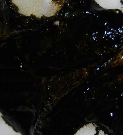
Black or Tarry Stools from Cancer vs. from Food or Drugs

Find out just what “tarry” stools look like when compared to black BMs from benign causes like food or medications.
How often have you read a pamphlet or online article about colon cancer and came upon the term, “tarry stools”? Tar? Have you even ever seen hot tar?
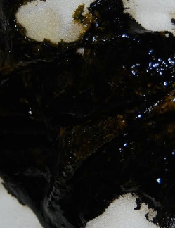
The reason that black stools are often referred to as “tarry” is because, says Michael Blume, MD, “Black stools from bleeding actually have a somewhat sticky appearance, much like tar, while this is often not the case if it is from foods or medications.”
Dr. Blume is a gastroenterologist at MedStar Good Samaritan Hospital, Baltimore.
“Tarry stools is a term that describes the appearance of the stool, where it looks sticky like tar.”

Tarry stools
Think back to a time when, perhaps, you saw what tar looked like on a sidewalk before it dried. Now, imagine that being mixed in with your BMs.
“We often show patients something black to make sure that they actually mean black.”
Are you absolutely sure that your bowel movements are the same hue as your raven dress shoes or coal-colored attire?
What else is “solid” black?
Place a stool sampling on a paper plate and hold it right beside a charcoal-colored item for comparison.
You might find that your poop is actually a very dark brown.
“Tarry stools usually relate to their visual description. In additional to being black, when from bleeding, they also can look rather sticky.” Sticky is the key word.
This all does not mean that if a disease process is causing very dark BMs, that they are necessarily tarry.
Dr. Blume says, “I certainly would not use appearance as the sole criteria with which to base a decision as to whether it was related to bleeding.
“Usually when one is bleeding from the upper GI tract enough to cause tarry stools, the stools are often looser and occurring more frequently.
“Blood is actually quite a good laxative, so one often looks at stool frequency and consistency as a measure of how actively someone is bleeding.
“Black stools that are formed and not frequent are often less likely to be related to bleeding.”
Very dark BMs can be caused by spinach, beets and Pepto Bismol, says Dr. Blume.
 In practice for 25+ years, Dr. Blume treats over 65 conditions including abdominal pain, appetite loss, blood in stool, celiac disease, colon cancer, esophageal and liver disease, gas and IBS.
 Lorra Garrick has been covering medical, fitness and cybersecurity topics for many years, having written thousands of articles for print magazines and websites, including as a ghostwriter. She’s also a former ACE-certified personal trainer.
.
Top image: Shutterstock/Photology1971
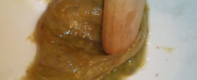
Benign Causes of Greasy Oily Stools
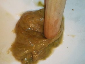
A GI doctor describes benign causes of oily or greasy bowel movements.
Do your stools look slimy, oily or greasy?
If they appear different lately, you should see a gastroenterologist, since only a doctor can determine for sure what the cause is.
“Greasy or oily stools, assuming one is not ingesting mineral oil, etc., is often a sign of fat malabsorption,” says Michael Blume, MD, a gastroenterologist at MedStar Good Samaritan Hospital, Baltimore.
“This can have many causes, including certain infections, as well as small bowel or pancreatic problems.
“Determining the cause often involves taking a detailed history as well as some diagnostic testing.”
What about IBS?
“One needs to differentiate oily looking stools and mucous in the stool,” says Dr. Blume.
“Many people with IBS complain of having mucous in their stools and that their stools look slimy.”
But mucous in the stools is not the issue with the conditions (described below) that cause oily bowel movements.
When stools are oily or greasy, this is called steatorrhea.
Pancreatic Problems
“One usually malabsorbs fat (this is what makes stools oily) if there is a problem with pancreatic function, such as with chronic pancreatitis, or from problems in the small intestine where fats get absorbed, such as with celiac disease, certain infections, such as Giardiasis, small bowel bacterial overgrowth, or other inflammatory diseases.”
One such inflammatory disease is Crohn’s disease.
Benign Causes of Greasy Oily Stools
Benign causes of steatorrhea, or oily stools, often involve dietary factors or non-serious gastrointestinal issues.
One common benign cause is the consumption of high-fat foods or supplements, such as those rich in oils or fats, which can lead to temporary steatorrhea.
You’ll be able to see the “greasiness” in your poops.
Another cause can be the use of certain medications, like those containing bile acid sequestrants, or specific weight loss drugs, which can interfere with fat absorption.
Additionally, rapid gastric emptying, also known as dumping syndrome, can lead to steatorrhea.
This occurs when food moves too quickly from the stomach to the small intestine, leading to insufficient digestion of fats.
Similarly, lactose intolerance can occasionally result in oily stools if the body can’t properly digest dairy products, though it more commonly causes diarrhea and gas.
These benign causes typically resolve with dietary adjustments or discontinuation of specific medications.
Can cancer cause oily stools?
Yes, cancer can lead to steatorrhea.
Pancreatic Cancer: This is one of the most common causes of steatorrhea.
The pancreas produces digestive enzymes that help break down fats.
If pancreatic cancer impairs enzyme production or blocks the pancreatic duct, it can lead to poor fat digestion and absorption, resulting in oily stools.
Biliary Tract Cancer: Cancers affecting the bile ducts or gallbladder can interfere with bile flow.
Since bile is essential for the digestion and absorption of fats, obstruction or disruption in bile flow due to cancer can lead to steatorrhea.
Small Bowel Cancer: Tumors in the small intestine can disrupt the digestive process and lead to malabsorption of nutrients, including fats, causing oily stools.
]In practice for 25+ years, Dr. Blume treats over 65 conditions including abdominal pain, appetite loss, blood in stool, celiac disease, colon cancer, esophageal and liver disease, gas and IBS.
Lorra Garrick has been covering medical, fitness and cybersecurity topics for many years, having written thousands of articles for print magazines and websites, including as a ghostwriter. She’s also a former ACE-certified personal trainer.
Blood Loss Symptoms of Black Blood in the Stools
“Black stools are often a sign of bleeding from a site in the upper GI tract, such as an ulcer, etc.,” says Michael Blume, MD, a gastroenterologist at MedStar Good Samaritan Hospital, Baltimore.
What else can make your bowel movements appear black?
“Common causes of this could include medications such as iron or Pepto Bismol,” says Dr. Blume, “as well as certain foods, such as spinach or beets.”
Black licorice has been known to turn bowel movements a very dark color. And even though spinach is green, a generous amount of this nutrient-dense vegetable can turn poops very dark.
Dr. Blume continues, “Symptoms are not usually the best guide, as many people who have bleeding ulcers often have no pain associated with their ulcer, and present with a complication of their ulcer, such as bleeding.
“Certainly, if one feels weak or dizzy, especially on standing up, it may be a sign that one is bleeding significantly.
“Additionally, one may have other signs of significant blood loss, such as cardiac symptoms and shortness of breath.
“Being anemic can impair your blood’s oxygen-carrying capacity and cause cardiac symptoms.”
Anemia can be caused by internal blood loss, such as that from an ulcer or a cancerous tumor in the colon.
Risk Factors for Bleeding Ulcers
Dr. Blume explains, “One may want to examine if they have risk factors for ulcers such as cigarette smoking, taking anti-arthritics, etc.
“One should be aware of the context in which one sees the black stools to best interpret this finding.”
Take note of what you eat.
If you see nearly black poops in the toilet bowl the day or two after you ate a lot of licorice, spinach, shellfish or organ meats (the last three are high in iron), that can very well be the explanation.
If there’s no correlation with food intake, see your doctor.
In practice for 25+ years, Dr. Blume treats over 65 conditions including abdominal pain, appetite loss, blood in stool, celiac disease, colon cancer, esophageal and liver disease, gas and IBS.
Lorra Garrick has been covering medical, fitness and cybersecurity topics for many years, having written thousands of articles for print magazines and websites, including as a ghostwriter. She’s also a former ACE-certified personal trainer.

Blood in Stools, No Other Symptoms: Colon Cancer?

Find out if bloody stools can be the only symptom of colon cancer.
How likely is it that if you see blood in your stools, but do not have any other symptoms (e.g., unexplained weight loss, abdominal pain), that you can actually have colon cancer?
“Colon cancer often presents with no symptoms,” says Michael Blume, MD, a gastroenterologist at MedStar Good Samaritan Hospital, Baltimore.
You’re probably wondering how such a patient’s colon cancer is then discovered if there are no symptoms.
It may be found incidentally, via imaging for an unrelated issue, or during a routine colonoscopy.
“Not all colon cancers bleed, and when they do bleed, it’s often is not visible, i.e., occult bleeding, which can be picked up on chemical tests, but not visible,” says Dr. Blume.
“This is why it is very important to have screening colonoscopies at the appropriate time, even if you have no symptoms. Remember that lack of symptoms does not mean lack of a problem.”
Blood in Bowel Movements and Colon Cancer
“Colon cancers usually do not cause tarry stools,” continues Dr. Blume.
“The bleeding that one sees is usually bright red or maroon. Bleeding may be the only sign that one may see, even with an advanced cancer.
“One can see pain, bowel dysfunction, weight loss, etc., with colon cancer.
“Usually, when one sees these symptoms when they are related to a colon cancer, it is often quite advanced.
“It is important to understand, however, that if one sees bleeding per rectum, the lack of other symptoms, such as pain, bowel dysfunction, weight loss, etc., does not mean that one does not have a cancer.”
- Blood in the stools — could be red, reddish brown or even black with a tarry look
- Pencil-thin or ribbon stools
- Feeling of incomplete voiding after a bowel movement
- Constipation or diarrhea
- Alternating constipation with diarrhea
- Odd change in bowel habits
- Abdominal pain or cramps, gas
- Feeling of bloating or fullness
- Unexplained weight loss or fatigue
- Back pain
In practice for 25+ years, Dr. Blume treats over 65 conditions including abdominal pain, appetite loss, blood in stool, celiac disease, colon cancer, esophageal and liver disease, gas and IBS.
Lorra Garrick has been covering medical, fitness and cybersecurity topics for many years, having written thousands of articles for print magazines and websites, including as a ghostwriter. She’s also a former ACE-certified personal trainer.
.
Top image: Freepik/yanalya

Can Menstruation Be Affected by Irritable Bowel Syndrome?

It’s not true that irritable bowel syndrome can affect menstruation.
“IBS, while a disorder that can cause rather inconvenient symptoms, is not a dangerous disease, and is not associated with any increased risk for other serious diseases,” says Michael Blume, MD, a gastroenterologist at MedStar Good Samaritan Hospital, Baltimore.
Irritable Bowel Syndrome and Your Period
“If one is having menstrual problems, it is usually caused by some other issue, not related, except perhaps peripherally, to IBS,” says Dr. Blume.
“What I mean is that the IBS itself is usually not the immediate cause of menstrual problems.
“It would suggest that there is some other issue going on that is not only causing the menstrual abnormalities, but also contributing to that person’s IBS-like symptoms.”
In short, two issues going on at once does not mean there is a causal relationship between the two.
There may be a third issue at hand, causing the first two issues.
For any suspicious gynecological or gastroenterological symptoms, always see the appropriate medical specialist.
The Reverse: Worsening of IBS Symptoms from Menstruation
IBS can become worse during menstruation – according to subjective reports by patients in a study by Whitehead et al.
However, the mechanism – if one even exists – of how a woman’s period can amplify her irritable bowel syndrome process is not known.
Cause and effect, or any kind of direct association between IBS and menstruation is challenging to decipher.
What makes the investigation into this so vexing is that irritable bowel syndrome and the monthly cycle have a symptom overlap.
For instance, the monthly cycle can cause diarrhea and lower abdominal cramps.
It’s also notorious for causing bloating. Some women report constipation as well.
A study in the British Medical Journal found that in women with IBS, menstruation increased rectal sensitivity.
The study authors believe that this suggests that patients with IBS respond differently to the fluctuations in hormones when compared to subjects without IBS.
In practice for 25+ years, Dr. Blume treats over 65 conditions including abdominal pain, appetite loss, blood in stool, celiac disease, colon cancer, esophageal and liver disease, gas and IBS.
Lorra Garrick has been covering medical, fitness and cybersecurity topics for many years, having written thousands of articles for print magazines and websites, including as a ghostwriter. She’s also a former ACE-certified personal trainer.
.
Top image: Shutterstock/Emily frost

Numb, Burning Foot with Intense Pain: Cause and Solution

Find out what might be causing the debilitating burning in your feet.
I fell in love with my husband on the dance floor. Yet, when I was diagnosed with type II diabetes at age 45, I disregarded the doctor’s advice and went on with my life like normal.
Since there wasn’t any pain associated with diabetes, I continued dancing with my husband, working and partaking in all day-to-day activities that for all intents and purposes created my diabetes.
It wasn’t until about 10 years later that my diabetes started to cause me pain. I can remember the night vividly:
I began feeling a sharp, piercing pain in my feet. This pain lasted the entire night and didn’t let up until later the next day.
Shortly thereafter, the excruciating pain became so persistent that I was forced to cut back on my work hours, forgo walks with my family and even give up dancing altogether. Just standing became unbearable due to the chronic pain.
Burning Feet
It felt as if someone was holding a blowtorch to my feet. The pain caused so many problems in my life, and the depression led to me shutting down and becoming a virtual hermit.
Diabetic neuropathy can cause “burning” and numb feet.
In 2007, I was diagnosed with diabetic neuropathy. Diabetic neuropathy is a nerve disorder resulting in irregular sensations that affect up to 70 percent of the 26 million Americans diagnosed with diabetes.
Diabetic neuropathy produces a range of conditions from pain so excruciating that sufferers can’t sleep with their feet under the bed sheets, to numbness so extreme that stepping on glass shards won’t be realized for weeks or even months.
Diabetic neuropathy made me completely dependent on my husband for all household chores.
Doctors would ask me to describe my pain level as a number from 1-10, but for me it was the distance between the earth and the moon.
I considered everything to alleviate the pain from higher doses of pain medication to a nerve-blocking spinal injection, but my endocrinologist questioned the success rate and risks.
He warned me that I would suffer from intense pain until the nerves in my feet died, which would eventually increase my risk of amputation.
I suffered for two years until my doctor put me on Metanx, a medical food pill that contains the active forms of folic acid, vitamin B6 and vitamin B12. I took the pill continuously for a few months but couldn’t tell if it was working … until one day I realized that I had gone days without remembering to take my pain medications!

Deborah Grona dancing with her husband
The end of burning feet.
Diabetic neuropathy changed my life, but Metanx helped to change it back.
Metanx is the only thing I can attribute to being pain-free, and I think it will play a huge part in helping those with diabetic neuropathy in the future.
I thought the life I once knew was gone indefinitely, but I’ve come out of the fog of pain and rejoined the world. And, most of all, I can dance with my husband again.
Top image: Shutterstock/Denis Val

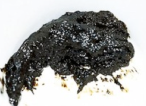
Can Beets Make Stools Almost Black?

Black stools can mean a serious medical problem, so how can you tell if this discoloration is from beets?
Have your stools appeared black lately, and did you wonder if this wasn’t from having recently eaten beets or the juice of this vegetable, and instead feel fearful that you might have a serious disease?
“Black stools are often a sign of bleeding from a site in the upper GI tract, such as an ulcer, etc.,” says Michael Blume, MD, a gastroenterologist at MedStar Good Samaritan Hospital, Baltimore.
“Other things can make your stool black, however,” says Dr. Blume.
“Common causes of this could include medications such as iron or Pepto Bismol, as well as certain foods, such as spinach or beets.”
So there you have it: Beets, indeed, can make bowel movements appear black, but be careful here.

Beents cannot make poops literally as solid black as chunks of coal.
However, in certain light, poops can appear off-black or an extremely dark brown, like a brownish-black.
What does this look like, when compared to the very dark discoloration from old blood?
Since I’m in that 10-15 percent of the population who experiences visible undigested beet pigment (betain), I can tell you what “black stool beeturia” looks like.
Whenever my BMs appear “black,” I’ve noticed that they are small and in clusters like big pebbles (size of marbles).
The color is actually off-black or a very dark brown. It’s possible that the size and shape of the poops create the illusion of a darker hue.
They aren’t this dark when they’re larger in diameter and more link-like.
Often, the smaller an object is, the deeper its color appears to be, at least when the color is inherently dark.
So what you are “seeing” as black bowel movements may actually be just a very dark brown.
I recommend removing a few from the toilet and setting on a paper plate in good light.
Then place something black, such an article of clothing, besides your poops and make the judgment.
The pigment of my darkest stools, resulting from beet ingestion, is actually an attractive brown-black. It has a smooth, rich quality and its uniform.
If the sight of “black” stools alarms you even though you know it has to be from the beets you recently consumed, you should increase your fiber intake—a lot.
This will loosen your bowel movements and tend to make them lighter, even if there’s betain in them.
In practice for 25+ years, Dr. Blume treats over 65 conditions including abdominal pain, appetite loss, blood in stool, celiac disease, colon cancer, esophageal and liver disease, gas and IBS.
Lorra Garrick has been covering medical, fitness and cybersecurity topics for many years, having written thousands of articles for print magazines and websites, including as a ghostwriter. She’s also a former ACE-certified personal trainer.
.
Top image: Gayvoronskaya_Yana

Black Stools from Colon Cancer vs. Acid Reflux

A doctor addresses the question of how do black stools from colon cancer compare to those caused by acid reflux.
I wanted to know if there were distinct differences in the appearance of black stools from acid reflux compared to those that can result from colon cancer.
“Black stools are not commonly seen with a problem in the lower gastrointestinal tract, as bleeding from the colon is more often bright red or maroon, assuming it is significant bleeding,” begins Michael Blume, MD, a gastroenterologist at MedStar Good Samaritan Hospital, Baltimore.
“Remember that colon cancers usually do not cause acute massive bleeding; they more often just ooze, so depending on the location, one may see some blood with a bowel movement or perhaps no visible blood at all.
“Blood is also a very good laxative, so a good way to determine if one is massively bleeding is to look at whether the stools are formed or loose, and how often they are occurring.
“Reflux disease in and of itself does not cause black stools, unless one is bleeding significantly from an ulceration related to severe reflux disease.
“If one is bleeding enough to cause black stools, one is usually having some significant bleeding.”
In practice for 25+ years, Dr. Blume treats over 65 conditions including abdominal pain, appetite loss, blood in stool, celiac disease, colon cancer, esophageal and liver disease, gas and IBS.
Lorra Garrick has been covering medical, fitness and cybersecurity topics for many years, having written thousands of articles for print magazines and websites, including as a ghostwriter. She’s also a former ACE-certified personal trainer.
.
Top image: Shutterstock/Twinsterphoto

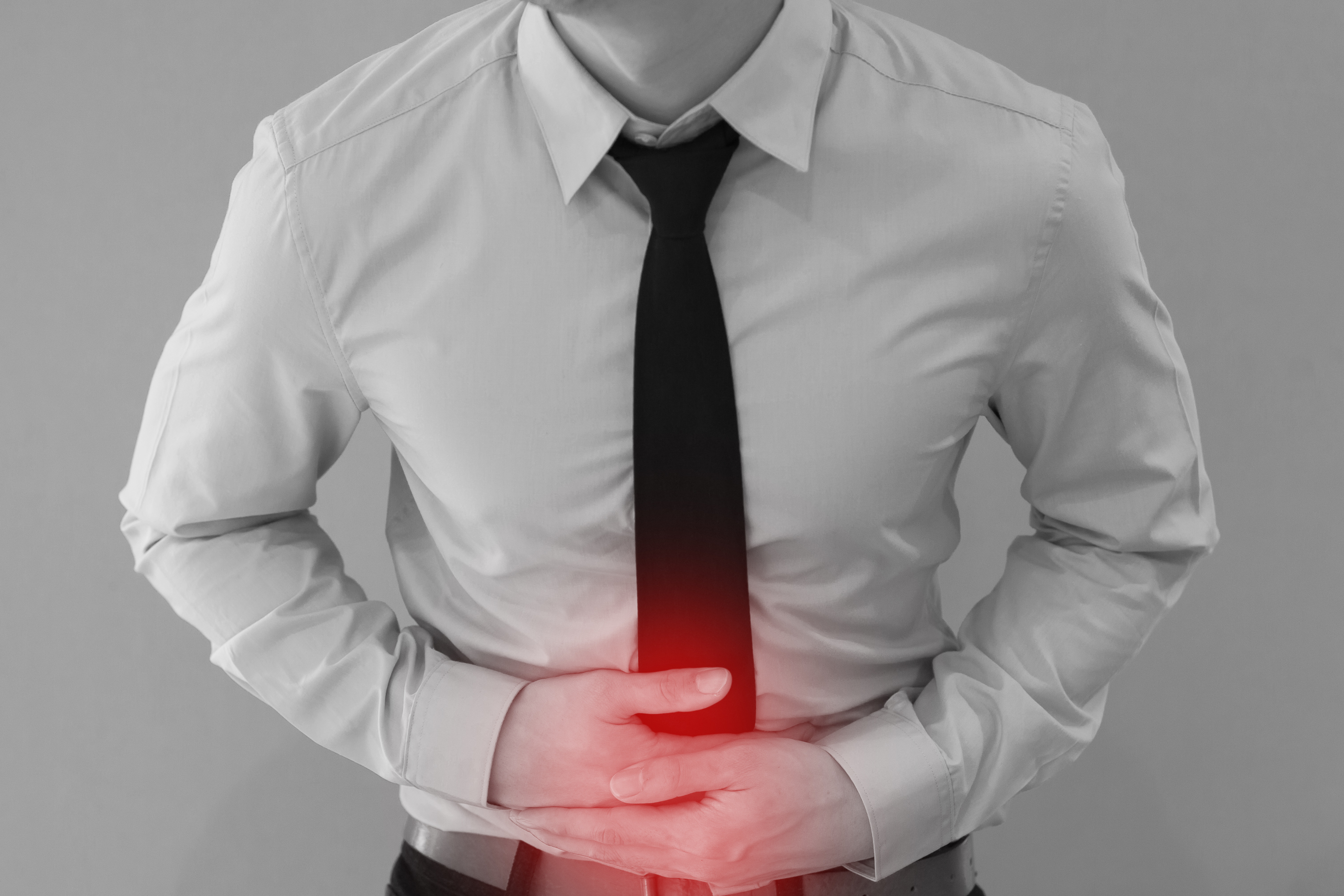
Causes of Nausea When Working Out in Well-Fed Athletes

There are several possible causes of nausea that occurs when you work out.
Are you troubled by the onset of nausea every time, or almost every time, you put in a good hard workout?
And I don’t necessarily mean after the training session, but during.
As a former personal trainer, I’ve had more than one client get “sick” during the course of a training session.
I can immediately recall three clients who got nauseated — two during the workout, and one immediately after.
All three were de-conditioned, and I worked them pretty hard.
One of them appeared to be under-nourished (underweight).
The intuitive explanation, then, for why a person suffers nausea during a workout is that they are undernourished, dehydrated and/or out of shape.
But what about fitter, well-fed, hydrated individuals?
“The causes of nausea while working out are likely similar to that of reflux in athletes,” says Steven Fleisher, MD, a gastroenterologist in Rosedale, Maryland, with 20+ years of experience.
“These include decreased emptying of the stomach due to diversion of blood flow away from the digestive system to the muscles.
“This may also affect contractions of the digestive system in general (dysmotility).
“Interestingly, it does not appear that stomach acidity is affected by exercise.
“However, increase in bile acids that can irritate the stomach and cause nausea have been reported.”

Dr. Fleisher was named a 2015-2018 “Top Doc” by Baltimore Magazine for gastroenterology.
Lorra Garrick has been covering medical, fitness and cybersecurity topics for many years, having written thousands of articles for print magazines and websites, including as a ghostwriter. She’s also a former ACE-certified personal trainer.
.


