

What Makes Gums Look White? Could It Be Cancer?

White gums are not normal. They mean something is wrong. This includes the possibility of cancer.
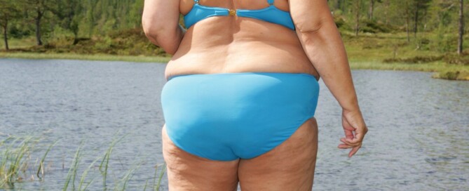
Should Plus Size Women Feel Pressure to Wear a Bikini?
Are you plus size and feeling pressure to wear a bikini or other “daring” clothes due to the body positivity movement?
Have influencers convinced you that this is the only way to show the world your confidence?
They’re called “influencers” for a reason. They want to influence the thinking of their followers and gain new followers to convert.
Have you been struggling with your weight for years and are now struggling with the guilt of betraying the body positivity movement because you STILL haven’t bought a bikini or worn some other type of daring clothes that big women aren’t “supposed to wear’?
It’s as though many influencers, including morbidly obese, are shouting, “Come ON already! Take the plunge! You’ll feel liberated! A bikini body is ANY body in a bikini! What’re you waiting for?!”

Shutterstock/DenisProduction.com
But despite all the hype, all the Instagram images, all the TikTok videos — about more and more very large women showing off their bodies in bikinis — you still just can’t get into this mindset.
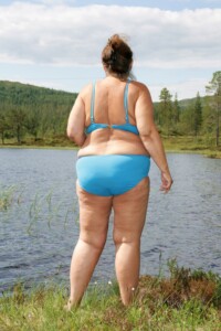
It’s Okay for Fat Women to Stay Covered up or Wear One-Piece Swimsuits at the Beach or Pool
You do not need to leap onto that bandwagon if the idea makes you feel uneasy or unnerved.
If you want to gain some serious empowerment, I suggest you pick up a barbell and start dripping sweat.
This is what I had my obese clients do when I was a fitness trainer.
They gained tremendous confidence knowing they were training their bodies to move heavy resistance, to climb flights of stairs with more ease, to do things that they never thought their body could do.
Since when has body positivity become a prescription for how much flesh you should show in public?
Would you buy shoes that were too small? No. Why buy a bikini if you KNOW you’ll feel uncomfortable in it?
Why not work on developing skills rather than struggling to work up the nerve to wear a bikini or crop top?
You’ll gain more empowerment and feelings of worthiness by doing any of the following:
Learning a second language, working on your speaking skills, learning to touch type, starting a garden, taking self-defense classes, learning to throw a punch, taking up yoga, volunteering for a favorite cause, learning about nutrition, spending more fun time with your kids … gee, the list is endless.
Stop worrying about the damn bikini. It’s just a prop.
Feeling Pressured to Wear a Bikini
If this is really bothering you, then you need to examine why you’ve allowed a complete stranger on Instagram to take you to this place.
Wear what feels comfortable to you. Where what feels like you.
For example, I myself would never be caught dead in a sun dress. The solution? I don’t wear sun dresses. Simple.
If you’d feel awkward, self-conscious or anything else negative in a two-piece swimsuit, or without a cover-up — then for Pete’s sake, keep wearing the one-piece with the cover-up. If for now, that’s what you feel at ease in, then DO IT.
Once you’ve made that decision, your anxiety levels will drop, and you’ll be able to focus on more important things in life than some prop.
It’s your legal and God-given right to hate wearing bikinis!
A full-figured woman has a right to be modest.
When you go to the beach or pool, you’re there to enjoy the environment, the water, play with your kids or supervise them.
You do not need to bare a lot of belly and thighs to do this.
My mother once told me, “If you have to talk yourself into buying a particular home, then it’s not the right one for you.”
Apply that to the bikini. If you have to talk yourself into wearing one in public — then the timing isn’t right for you. Ditch the idea and enjoy life.
 Lorra Garrick is a former personal trainer certified through the American Council on Exercise. At Bally Total Fitness she trained women and men of all ages for fat loss, muscle building, fitness and improved health.
.
Top image: Inger Anne Hulbækdal

Seeking Chronic Pain Treatment? What Your Doctors Missed

People suffering with low back pain in the morning, neck pain or sciatica when sitting in a chair, hip pain when walking, shoulder pain or any other musculoskeletal condition…
…are still pursuing the same treatments they were seeking decades ago: (more…)

10 Things You’d Never Think Would Sabotage Weight Loss Efforts

Have you been struggling to lose weight but it just won’t come off? There are reasons for this – explanations you’d never even think could be responsible.

Causes of Numbness on the Outside of the Big Toe

Are you scared that the numbness on only the outside of your big toe is an early symptom of MS, diabetes or some other serious illness? (more…)

Percentage of a Surgeon’s Lung Cancer Patients Who Smoked

Many smokers never get lung cancer, but then again, most lung cancer patients are smokers.
Furthermore, if smokers lived longer, it’s fair to wonder how many would have eventually developed lung cancer. (more…)

Can You Be Too Young to Get Multiple Sclerosis?

There’s a reason you should never think you, or your child, is too young to develop multiple sclerosis, an autoimmune disease that can cause severe disability and a host of symptoms. (more…)

Late Onset Multiple Sclerosis vs. Early Onset Symptoms?

Is there a difference between the symptoms of late onset MS and early onset MS? (more…)

Is There a Way to Live Longer with Multiple Sclerosis?

If you’ve been diagnosed with multiple sclerosis, you have more control over how long you live than you may think.
There ARE ways to maximize your life expectancy even though you have MS. (more…)
Causes of Tingling, Burning in Both Feet Including Neurological

The key word is “both” feet. Unfortunately, your fear of diabetes or a spinal tumor as the cause of burning and tingling is realistic.
But most causes of this symptom pair are certainly not from a tumor (more…)


