
What if You Get a Pulmonary Embolism on a Plane?

Suppose a passenger on an airplane develops an apparent pulmonary embolism; what should, or CAN, you do?
A deep vein thrombosis can develop while a person is on an airplane.
What might be done for help if this DVT migrates and becomes a pulmonary embolism, and a doctor just happens to be on the plane—a doctor who has treated pulmonary emboli?
“You will not show signs of a pulmonary embolus on an airplane,” says Michael P. Zimring, MD, Director, Travel Medicine, LLC at Mercy Medical Center, Baltimore, Maryland..
“The PE usually breaks off from a clot starting in the lower extremities — and that takes a while to develop.
“You usually show signs of a pulmonary embolus after a significant time of first forming a clot in the leg. Sometimes it is fast, but not usually.”
In a hospital setting, the treatment for a pulmonary embolus is IV administration of a “clot busting” or thrombolytic drug.
Doctor on Airplane Suspects Passenger Has a Pulmonary Embolism
But what if a doctor on a plane suspected that a passenger had a PE (based on symptoms, risk factors), and that doctor just happened to have a clot busting drug and syringe in his medical kit?
These are fair questions for a layperson to wonder about.
Though thrombolytics carry the risk of dangerous internal bleeding as a side effect, it’s reasonable for a layperson to wonder about this if a passenger has an apparent pulmonary embolism and is 35,000 feet off the ground.
Diagnosing a Pulmonary Embolism
A pulmonary embolism cannot be confirmed based on just symptoms (sudden difficulty breathing, chest pain, coughing up blood) and risk factors for DVT (prolonged inertia in an airplane, obesity, smoker, among many others).
Back to that doctor on the plane. What if he also has a stethoscope and listens to the distressed airline passenger’s heart?
A pulmonary embolism can change the way the heartbeat sounds through a stethoscope.
Dr. Zimring explains, “A typical physician will not hear a strain of the right heart through a stethoscope, and no one will be able to detect a PE by physical means.
“No one would dare treat a pulmonary embolus by injecting a thrombolytic on a plane because there is no way he could be sure the patient has a PE.
“There are many factors to be taken in consideration before injecting a thrombolytic in the best hospital environment.
“In addition, the physician might not be covered under the Good Samaritan Laws if he did such a specialized procedure in an emergency situation.”
Prevention of Blood Clots Triggered by Air Travel
If you’re worried about developing a pulmonary embolism as a result of flying, take precautions:
Stay well-hydrated throughout the plane trip; exercise the calve muscles every 15 minutes during the flight; don’t be seated for more than an hour; and avoid alcohol.

Exercises during air travel
 Dr. Zimring has 40+ years of experience diagnosing and treating a host of medical conditions.
 Lorra Garrick has been covering medical, fitness and cybersecurity topics for many years, having written thousands of articles for print magazines and websites, including as a ghostwriter. She’s also a former ACE-certified personal trainer.
.
Top image: ©Lorra Garrick

Quick Easy Way to Tell if Red in Stools Is Beet Juice

Here’s how to tell within seconds if the “blood” you see in your bowel movements is actually undigested beet juice.
I juice beets all the time, and I experience “stool beeturia” as a result.
Perhaps the biggest telltale sign that the red in your poops is from the beet pigment betain, is that it has a glowing, neon-like quality.
Even veteran beet eaters can still wonder if the red in their stools is blood, especially if they’ve recently had a digestive problem.
This is the same glowing, neon-like quality you see when a glass fills with the juice of a fresh beet being juiced.
Pictures of glasses of beet juice on the Internet don’t do this pigment justice.

Shutterstock/bitt24
Most website images of beet juice in glasses seem to be beet juice blends.
Read the descriptions; almost always, there are other items juiced into the beverage, such as carrots, blueberries, apples, celery or some leafy green.
This suppresses that glowering, neon-like color. If you juice ONLY beets, you’ll be struck by the amazing crimson glare.
It’s beautiful and bright. This quality is retained in your stools when betain does not get absorbed.
Blood in the stools does not look like this.
Though blood in the stools may be fresh (and “bright red”), it lacks that glow, that neon-like feature, that distinct ruby-magenta or crimson-magenta color, whereas blood is simply a solid red.
There’s a difference between crimson-magenta and solid red.
The exact color of betain in BMs seems to be 80 percent crimson, 20 percent magenta, with neon. This hardly describes blood, fresh or old.
As a woman, I’ve seen blood in the toilet with (NOT in) my stools as a result of menstruation.
If blood is being discharged from my vagina at the same time I’m having a bowel movement, the blood will sometimes get “mixed” with the poops.
I know what this looks like all too well. And it cannot pass for betain. Quite simply, betain often just GLOWS.

Freepik.com
 Lorra Garrick has been covering medical, fitness and cybersecurity topics for many years, having written thousands of articles for print magazines and websites, including as a ghostwriter. She’s also a former ACE-certified personal trainer.
.
Top image: Shutterstock/KarepaStock

How Many Burps per Minute from Anxiety? How to Stop?

Wait till you find out how many times per minute anxiety can make a person burp.
It’s no secret that anxiety can cause burping, but just how much?
“In extreme cases, belching can occur up to 20 times a minute,” says Dr. David Robbins, a gastroenterologist at the Manhattan Endoscopy Center, NY.
“But in these cases it’s usually release of air from the esophagus back up into the pharynx without ever getting down into the stomach.”
The sound of a belch occurs when air escapes from the esophagus into the pharynx, says Dr. Robbins.
But he adds, “The most common type of belching originates when swallowed air from the esophagus enters the stomach, only to be released when the valve at the bottom of the esophagus relaxes.”
He refers to the type of burping, in which the released air was never in the stomach (only in the esophagus) as “supragastric.”
Most patients who have this type of burping “suffer from some element of anxiety,” continues Dr. Robbins.
“Studies have shown that the symptoms increase along with increasing stress. It’s also been described with obsessive-compulsive disorder and various eating disorders.”
So how does one stop burping 20 times a minute?

Shutterstock/Aaron Amat
“Interestingly, many patients can ‘squelch the belch’ while speaking,” says Dr. Robbins.
“This is basically a sort of distraction maneuver. This type of belching never occurs during sleep, again pointing to an active anxiety component.”
If you find yourself burping many times in just one minute, talking incessantly to squelch it isn’t practical, for obvious reasons.
What else can you do, then, besides making efforts to manage stress?
“There are no established treatment regimens for excessive belching,” says Dr. Robbins.
However, he adds, “Extreme cases require the help of a cognitive behavioral psychologist or a psychiatrist. Speech therapy and biofeedback have shown some modest results, while drugs such as simethicone (or Gas-x) have not.”
When circumstances are normal, says Dr. Robbins, burping “actually occurs up to 30 times a day” and is a “normal physiologic phenomenon,” even though we all have known at least one individual who can emit loud belches seemingly at will.
 Dr. Robbins is board certified in both gastroenterology and internal medicine. He has been invited to speak internationally and has published and lectured widely.
Lorra Garrick has been covering medical, fitness and cybersecurity topics for many years, having written thousands of articles for print magazines and websites, including as a ghostwriter. She’s also a former ACE-certified personal trainer.
.
Top image: ©Lorra Garrick

How Many People in Their Mid 30s Get Colon Cancer?

People in their mid 30s get colon cancer.
Many doctors don’t first suspect this in a symptomatic 30ish person, and the disease thus progresses until it’s finally diagnosed.
At the age of 35, the last thing Dr. Stan Frager was worried about was colon cancer.
On family vacation, Stan’s dad noticed that Stan had bleeding in his urine, and recommended that he see a doctor.
Upon returning from vacation, his father called Stan every day until he got screened.
35 too Young?
Stan says, “I only agreed to be examined to get him off my back.” Up until that point, he had been ignoring the bleeding in his urine, figuring it would go away.
But following a biopsy, a doctor informed Stan he had stage three colon cancer; his colon wall was penetrated, and he had a 90% morbidity rate.
“My father saved my life,” says Stan. Following a surgery, he went into stage 5 radiation treatments — the highest treatment available.
Before his diagnosis at age 35 for the colon cancer, Stan was an athlete and the baseball coach at the University of Louisville, Kentucky, where he was also a professor of psychology.
He had spent years in a medical environment and knew his way around the hospital, but never thought to get screened for colon cancer.
After his surgery he spent the rest of the baseball season coaching the team from the dugout, where he sat on a doughnut while he healed.
Despite the grim diagnosis, Stan has been a survivor for 38 years as of March 6, his surgery date.
He now has two full grown children, one of whom is a 28-year-old male who has already had a colonoscopy.
Stan hopes that audiences will take two simple messages away from his story:
- Know the symptoms and signs of colon cancer.
- Get checked out if something doesn’t seem right.
Age 35 is not too young to develop colon cancer.
“I spent 12 years in academia, and I was stupid. My, and many others, most common thoughts are ‘Why me? Maybe it’ll go away. I won’t get cancer.’ Then I did,” says Stan.
“This could have been prevented if I just took my head out of the sand.”
To learn more about this unforgiving disease, visit the Colon Cancer Prevention Project.
How often do people in their 30s get colon cancer?
Of all the cases in the U.S., the percentage of colon cancer patients between 20 and 34 is 1.4 percent; and between 35 and 44 is 4.2 percent.
There are no statistics that precisely define how many people in their 30s each year are diagnosed with colon cancer.
 Update: Stan Frager passed in 2021.
Top image: Shutterstock/Antonio Guillem
Source: seer.cancer.gov/statfacts/html/colorect.html
How Often Women with Dense Breasts Should Get Mammogram
Women with dense breasts have a significantly increased risk of breast cancer. Is the yearly mammogram often enough for adequate for tumor detection?
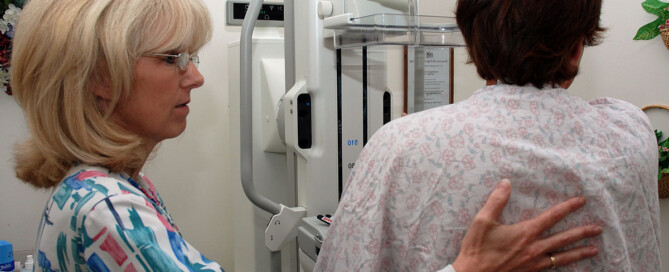
Because dense tissue increases the risk of breast cancer, one may wonder if a woman with dense breasts should have mammograms more often than women without this feature.
“Annual mammogram starting at the age of 40 saves the most lives and is appropriate for women of all breast densities,” says Anjali Malik, MD, a board certified diagnostic radiologist with Washington Radiology in Washington, DC. She interprets mammograms, breast MRIs and ultrasounds, and performs biopsies.
“Breast tomosynthesis [3D mammogram] has improved the detection of small, invasive breast cancers and decreased the ‘false positive’ call-backs,” continues Dr. Malik.
A report in JAMA Internal Medicine (March 2013) says that a study involving over 900,000 women concludes that mammograms once every two years are just as beneficial as annual screenings—even for patients with dense breasts.
The every-two-year screening results in fewer false positives, meaning, fewer indications of malignancy that turn out to be benign.
The researchers (UC San Francisco and Seattle-based Group Health Research Institute) say these recommendations apply to women 50 to 74.
Dr. Malik explains, “In 2013 (the time of the JAMA report), breast tomosynthesis (3D mammography) was not in routine use.
“The 2013 JAMA report was based on 2D analog mammography and prospective data from 1994-2008.
“Currently, 3D digital mammography is in use, and most centers use fellowship trained breast imaging radiologists, where this was not as widespread in the ‘90s and early 2000s. Therefore, this data is near obsolete in 2019.”
What about women in their 40s with dense breasts?
“For women, particularly for those women ages 40-49, who are also more likely to have dense breast parenchyma, nine per 10,000 breast cancer deaths are prevented by annual screening mammograms,” says Dr. Malik.
This data comes from a study by Nelson et al (Annals of Internal Medicine, 164(4):244-55, 2016).
A woman with dense breasts may wonder if more of an important screening tool means better: twice a year mammograms, whether they’re 3D or 2D.
But here’s the problem (other than the extra radiation exposure): Biannual screening increases the call-back rate and the rate of false positives.
This creates tremendous anxiety and sometimes unnecessary biopsies.
Though biannual screening via mammogram has been shown by limited research to result in more cancers detected, it has not been shown to increase survival rates.
Women with dense breasts (as determined mammographically) should ask their doctor about annual 3D mammograms supplemented with annual ultrasounds.
 Dr. Malik is a frequent public speaker and advocate for breast health awareness. She has lectured on the latest advances in breast cancer screening including 3D Mammography™. Follow her on Instagram: @AnjaliMalikMD
Lorra Garrick has been covering medical, fitness and cybersecurity topics for many years, having written thousands of articles for print magazines and websites, including as a ghostwriter. She’s also a former ACE-certified personal trainer.
.
Source: sciencedaily.com/releases/2013/03/130319144537.htm

Does Back Pain with Chest Pain Mean a Heart Attack?

What are the odds that if you uexplainably feel pain in both your back and your chest that it’s a heart attack?
Perhaps you’ve read that back pain can signal an oncoming heart attack, or occur with the actual heart attack.
Maybe you’ve heard that both back and chest pain can occur with a heart attack.
However, does pain in the back, plus chest, automatically mean that there is something wrong with your heart?
“Not necessarily, but possibly,” says Robert M. Davidson, MD, a cardiologist with SignatureMD.
“If it is associated with exertion or stress, it might be heart related. If it is affected by movement or position, it is more likely to be muscular-skeletal.”
A condition that can cause back and chest pain, upon body position or movement, is costochondritis, an injury to the cartilage in the ribcage. This can hurt pretty bad, but it is benign.
Dr. Davidson continues, “Severe back pain, sometimes radiating to the chest, can be a sign of a leaking or ruptured aortic aneurysm, which is a medical emergency, and should be considered if someone has risk factors such as high blood pressure, older age, or known arteriosclerosis. The type of pain associated with this is often described as tearing.”
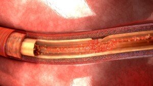
A tear in the inner lining of the aorta. Shutterstock
People who have survived a ruptured aortic aneurysm, or aortic dissection, have also described the feeling as “ripping.”
An aneurysm is an abnormally dilated or enlarged section of an artery.
Usually there are no symptoms until the aneurysm tears. Most aortic aneurysms are discovered by accident via imaging for another issue.
Most non-leaking or non-rupturing aneurysms don’t cause symptoms, but when they do, they may consist of chest, back, neck and jaw pain, a hoarse voice, abnormal stethoscope sounds, coughing and shortness of breath.
An esophageal spasm is another possible cause of chest pain that radiates to the back (including between the shoulder blades).
This is a benign (though sometimes very painful) non-cardiac issue and has no known cause.
Keep in mind that you can have both a musculoskeletal problem and a heart problem — both causing chest and back pain!

Dr. Davidson is with the Division of Cardiology at Cedars-Sinai Medical Center, Los Angeles, and has been practicing for 35+ years. Areas of specialty include coronary artery disease, heart attack and palpitations.
Lorra Garrick has been covering medical, fitness and cybersecurity topics for many years, having written thousands of articles for print magazines and websites, including as a ghostwriter. She’s also a former ACE-certified personal trainer.
.
Top image: Shutterstock/ArtFamily
Source: webmd.com/digestive-disorders/tc/esophageal-spasm-topic-overview

How Are Kids and Teens Treated for Blood Clots?

Yes, children, teens and young adults can develop a potentially fatal blood clot, also known as a deep vein thrombosis (DVT).
Even fit kids, under the wrong circumstances, can end up with this serious condition that demands immediate treatment.
Blood clots in kids, as well as in adults, pose a serious risk because they can break loose and travel to the lungs, resulting in a potentially life-threatening condition known as a pulmonary embolism.
When a blood clot lodges in the lungs, it obstructs blood flow, which can lead to sudden and severe symptoms such as difficulty breathing and chest pain.
A pulmonary embolism may cause rapid onset of shortness of breath, sharp or stabbing chest pain, and in some cases, even coughing up blood.
Immediate medical attention is crucial for managing a pulmonary embolism, as prompt treatment can significantly impact the outcome and reduce the risk of severe complications or death.
Young adults and children are not immune to DVT , which is a blood clot in a vein.
DVT stands for deep vein thrombosis. It’s a clump of blood — a sticky thick network of blood cells — attached to the inner wall of a vein, obstructing blood flow.
“Kids and teens are treated for blood clots according by identifying the underlying cause of the blood clot,” says Dr. Lisa Lewis, MD, a board certified pediatrician in Fort Worth, Texas, and author of “Feed the Baby Hummus, Pediatrician-Backed Secrets from Cultures Around the World.”
“Typically, blood clots are treated with an anticoagulant [blood thinner] and sometimes aspirin.”
Blood thinners would include heparin, or its low molecular weight version, given with warfarin (Coumadin), or just warfarin alone.
The INR blood work is taken on a scheduled basis so that the effect of the anticoagulant drug can be monitored to ensure it stays within therapeutic range.
“Although uncommon, a large percentage of blood clots in children and teens are inherited,” says Dr. Lewis.
What if a child or young adult has an acute, large DVT?
A large deep vein thrombosis can completely obstruct flow of blood from a limb.
In this case, other medications may be used early on, such as a thrombolytic (clot-busting) agent. This would be followed by anticoagulant medications.
Long-term management of deep vein thrombosis (and pulmonary embolism) in kids:
1) The safe prevention of additional DVTs is emphasized
2) Management is designed to enable the young patient to function as normally as possible.

Having 25+ years’ experience, Dr. Lewis completed her pediatrics residency at Texas A&M University Health Science Center, Scott and White Memorial Hospital. For two years afterward she was assistant professor in the department of pediatrics at Texas A&M University Health Science Center.
Lorra Garrick has been covering medical, fitness and cybersecurity topics for many years, having written thousands of articles for print magazines and websites, including as a ghostwriter. She’s also a former ACE-certified personal trainer.
Source: The National Alliance for Thrombosis and Thrombophilia newsletter at stoptheclot.org/Newsletters/NATT_Spring_07_Nwsl__web.pdf

12 Causes of Blood Clots and DVTs in Children

There are at least 12 causes of blood clots or deep vein thromboses in children. No age is immune.
Blood clots can occur anywhere in the body, but when they occur in larger vessels, they can become a serious problem.
When it comes to any kind of blood clot anywhere in the body of a child, the list of causes grows.
Not all blood clots are a deep vein thrombosis.
For example, a blood clot can occur in the brain due to head trauma.
There are causes of blood clots and deep vein thrombosis in the pediatric population.
“Genetic clotting disorders are very common,” begins Dr. Lisa Lewis, MD, a board certified pediatrician in Fort Worth, Texas, and author of “Feed the Baby Hummus, Pediatrician-Backed Secrets from Cultures Around the World.”
“Other causes include certain medications, surgery, anomalies of the blood vessels (for example a blood vessel that is not developed properly), immune system disorders, heart disease, immobility due to a medical condition or hospitalization, injuries, infection and cancer.”
Poor Blood Circulation
This can result from excessive inertia or constriction of a vein. Excessive inertia can easily be remedied by imposing limitations on how much time is spent watching TV and sitting around with a smartphone or computer game.
Turn the TV off and tell your child to go outside and play. HOWEVER, the directive of “go outside and play” is often not enough to encourage physical activity in a sedentary child.
But at least squatting down and making mud patties engages more muscles than does slumping on a couch staring at a screen.
Make sure your child has ready access to items that encourage movement: various balls, skates and other implements.
This can even include an air-filled striking bag for kids to have fun punching and kicking at.
A mini trampoline is also an option for older kids to offset inertia — and hence, reduce the risk of blood clots.
Damage to a Vein’s Inner Lining
Catheter placement into a vein can damage the inner lining of the vessel. Certain medications can also have this effect.
When children present with a DVT or pulmonary embolus, a combination of the above potential causes is often present.
Birth Control Pills
“Adolescents on birth control pills are more at risk of clotting disorders,” says Dr. Lewis.
“This is enhanced by smoking. Smoking is an independent risk factor for the formation of a deep vein thrombosis. So is obesity.
“At times a blood clot forms in a child for no apparent reason,” adds Dr. Lewis.
“A blood clot forms for various reasons depending on the cause. In general, a clot forms because of a cause that activates the components of clotting.
“For example, children with genetic clotting disorders may be missing a protein necessary for normal blood flow.”
Having 25+ years’ experience, Dr. Lewis completed her pediatrics residency at Texas A&M University Health Science Center, Scott and White Memorial Hospital. For two years afterward she was assistant professor in the department of pediatrics at Texas A&M University Health Science Center.
Lorra Garrick has been covering medical, fitness and cybersecurity topics for many years, having written thousands of articles for print magazines and websites, including as a ghostwriter. She’s also a former ACE-certified personal trainer.
.
Top image: ©Lorra Garrick

Who Should Have Upper Endoscopy to Diagnose Heartburn?

Too many upper endoscopies are being done unnecessarily, so if you have heartburn (GERD), take notice of this information.
If you’ve been diagnosed with heartburn secondary to GERD, but you have not had an upper endoscopy, have you ever wondered if you need this procedure?
A person may also find themselves tinkering with the idea of an upper endoscopy after learning that years of heartburn can lead to precancerous changes in the esophagus (Barrett’s esophagus).
“GERD or gastroesophageal reflux disease is a very common condition, with heartburn being the most common symptom of GERD — and seen in up to 50% people with GERD,” says Alan Gingold, DO, a board certified gastroenterologist with Central Jersey Ambulatory Surgical Center.
“Not everyone with heartburn needs to get an upper endoscopy,” continues Dr. Gingold.
“Typically an EGD is recommended for anyone who has heartburn that is longstanding (more than five years), symptoms not responding to medication, anyone aged 45 or over who has new symptoms or anyone with alarm symptoms: nausea/vomiting, dysphagia, anemia, blood on stool, weight loss.”
Dysphagia is difficulty swallowing.
Nevertheless, there may still be too many unnecessary endoscopies being done. In 2012, the Annals of Internal Medicine reported this.
Though that was years ago, it’s quite possible that people with heartburn are continuing to get unneeded upper endoscopies.
- After all, ads for heartburn relief continue to regularly air on TV and appear in print media.
- Furthermore, Barrett’s esophagus has received increased publicity.
However, if you seem to have heartburn or even if you were recently diagnosed with this condition, the next step is NOT to gun for the upper endoscopy.
Other Reasons for an Upper Endoscopy
If one’s heartburn doesn’t respond to medication after 4-8 weeks, this may call for an upper endoscopy.
Another reason for this exam is if the patient’s esophagus has a history of tightening or narrowing.
If the patient is over 50, male, has had diagnosed heartburn for at least five years, AND has other risk factors for Barrett’s esophagus – then he’s a candidate for an upper endoscopy.
Other Risk Factors for Barrett’s Esophagus
- Overnight reflux symptoms
- Excess body fat, especially in the belly
- Hiatal hernia
- Tobacco use
This patient may undergo an initial screening test for Barrett’s esophagus or esophageal cancer. If the result is negative, then periodic upper endoscopy is not needed.
What if you have Barrett’s esophagus?
Upper endoscopy should be done every three to five years.
If the patient has dysplasia, the upper endoscopy should be more frequent.
Overuse of upper endoscopy has not been found to improve the health or survival of patients suffering from heartburn.
 Dr. Gingold attributes his success to the extra time he spends with his patients. His areas of expertise include reflux disease, Barrett’s esophagus, capsule endoscopy, chronic liver disease and inflammatory bowel disease. Dr. Alan Gingold is board certified by the American Board of Internal Medicine in Gastroenterology
Lorra Garrick has been covering medical, fitness and cybersecurity topics for many years, having written thousands of articles for print magazines and websites, including as a ghostwriter. She’s also a former ACE-certified personal trainer.
.
Top image: Shutterstock/airdone
Source: sciencedaily.com/releases/2012/12/121203183341.htm

What’s the Strongest Risk Factor for a Deep Vein Thrombosis?

You’ll be shocked at what the greatest risk factor is for a DVT (deep vein thrombosis).
If you’re thinking that obesity is the strongest risk factor for a DVT — you’re on the right track as far as the seriousness of this risk factor.
But there’s another one that trumps obesity, says a study.
And that biggest risk factor is the non-O ABO blood type, according to a study that’s published in the Canadian Medical Association Journal.
The report says that this blood type comprises up to 20 percent of risks for VTE (venous thromboembolism).
Data on over 66,000 people, who’d been tracked for 33 years, was analyzed.
But could the ABO blood type be linked to a higher risk of DVT in the general population?
The study authors determined that DVT risk grew when the ABO blood type was combined with the factor V Leiden R506Q or prothrombin G20210A, which are genetic mutations that increase venous thromboembolism risk.
The researchers add that ABO blood type was indeed the most important risk factor in the general population for DVT/PE.
Other Big Risk Factors for Deep Vein Thrombosis
Obesity. “There are probably multiple factors relating to obesity and DVT risk,” says Susan L. Besser, MD, with Mercy Medical Center, Baltimore; Diplomate, American Board of Obesity Medicine and board certified by the American Board of Family Medicine.

Freepik.com
Dr. Besser explains, “It is theorized that significant obesity increases the hyper-coagulability of the blood (tendency to clot), especially if the person has a genetic predisposition to clotting (certain blood factors that are inherited can affect a person’s clotting ability).
“Additionally, physical factors, such as immobility (common in the obese) is another factor.”
Smoking. Quit. Just quit. Find another way to deal with stress or boredom.
Excessive sitting. Do you have a sit-down job? Every hour, get up and walk somewhere briskly, or do stationary lunges, squats or high knee marches for several minutes.
No exercise program. Start lifting weights and using cardio equipment, or join a cardio class. Go on hikes, bike rides, etc.
Though the greatest risk factor for a deep vein thrombosis is a blood type that you may not even have, this doesn’t mean that the other risk factors are insignificant. They count big-time.
-
- Abdominal surgery — recent
- Age over 60
- Airline travel — prolonged
- Bedrest — extended
- Birth control pills
- Cancer (active malignancy)
- Chemotherapy
- Chronic heart failure
- Chronic kidney disease
- Chronic venous insufficiency
- Dehydration
- Factor V Leiden mutation
- Family or personal history of DVT
- Fracture, major trauma
- Heart attack
- Hormone replacement therapy
- Joint replacement surgery: recent
- Obesity or moderate overweight
- Paraplegia or quadriplegia
- Pregnancy — current
- Sedentary lifestyle
- Sitting excessively most days
- Smoking, chewing tobacco
- Ultra-processed food diet and lastly, varicose veins