
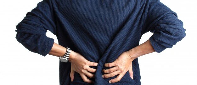
Low Back Pain Cure with Simple Walking on a Treadmill
A consistent walking program using a treadmill can dramatically reduce or even eliminate low back pain. But if you use a treadmill, there’s a catch.
A report in Clinical Rehabilitation presents conclusions of a walking-study done with participants who had low back pain.
Frequency and Duration of Walking to Effectively Treat Low Back Pain
The program in this study, headed by Dr. Michal Katz-Leurer from Tel Aviv University’s Stanley Steyer School of Health Professions (Sackler Faculty of Medicine), consisted of walking two or three times a week for 20 to 40 minutes at a time.
This walking protocol turned out to be as effective for relieving low back pain as are the typical muscle strengthening exercises that require equipment and often supervision by rehab specialists.
How does walking cure or relieve low back pain?
Few people actually realize that the “core” is involved in walking. I’m a former certified personal trainer, and I’ve told my clients that the core—which is primarily the abdominals and low back—is engaged when one walks.
This is why when some very sedentary people suddenly do a lot of walking, their low back starts aching. These muscles have worked in a way they’re not used to.
Your body doesn’t have to bend in order to engage the core.
However, don’t let the possibility of backache stop you from a regular walking program if you suffer from low back pain.
You may feel some achiness at first, but it will subside sooner than you think as you stick with the program.
It’s like any new exercise to the body; the muscles are adjusting to the new stimulus, and in the process, develop benign aching or soreness. As the muscles get stronger or adapt, they will no longer get sore.
How the Study Was Done
Twenty-six people completed a muscle exercise program, and 26 others completed a walking program.
All participants had low back pain, and both programs lasted six weeks.
At the end of six weeks, both groups had great improvement in their condition.
How to Properly Use a Treadmill to Alleviate Back Pain
In order for walking to effectively treat low back pain, it must be done with correct biomechanics.
This means if you use a treadmill, DO NOT HOLD ON — other than for momentary steadying such as when drinking water.

Shutterstock/Khakimullin Aleksandr
Otherwise, when your hands are latched onto the sides or front, even lightly, this disrupts the natural gait pattern and can make low back pain worse, as well as cause new pains elsewhere in the body, such as in the hips, knees and feet.
If your back hurts more upon walking on a treadmill, this is because your lumbar muscles are being engaged. This achiness will especially occur if you use an incline without holding on.
This is because when you walk on a slope or incline, without holding on, your lower back muscles work to prevent you from falling backwards!
If you hold on, this will relieve the soreness or aching, but holding onto the treadmill is WRONG!
Keep walking without holding on, and that aching in your low back will vanish. Give those weak, out of shape muscles a chance to get strong and reliable!
Use a SLOW pace and medium to low incline. There is no need to crank the machine up to 15 percent and run the tread at 4 mph, which is too difficult to sustain a walk without holding on.
But holding onto a treadmill at ANY setting will cheat your low back from the work it needs to help prevent or eliminate pains and aches.
 Lorra Garrick is a former personal trainer certified through the American Council on Exercise. At Bally Total Fitness she trained women and men of all ages for fat loss, muscle building, fitness and improved health.
.
Top image: Freepik.com/shayne_ch13
Source: sciencedaily.com/releases/2013/03/130305131404.htm
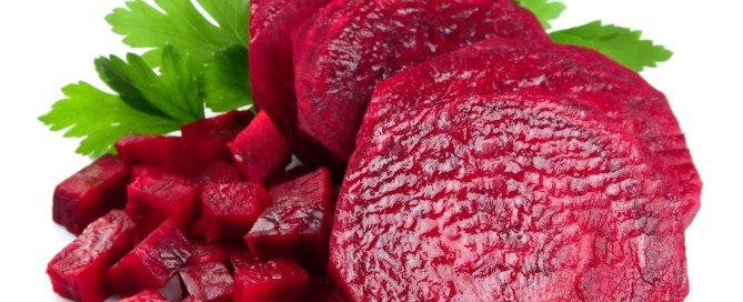
How Long Can “Stool Beeturia” Last?
Here is what a GI doctor says about how long beet juice in one’s stools can last.
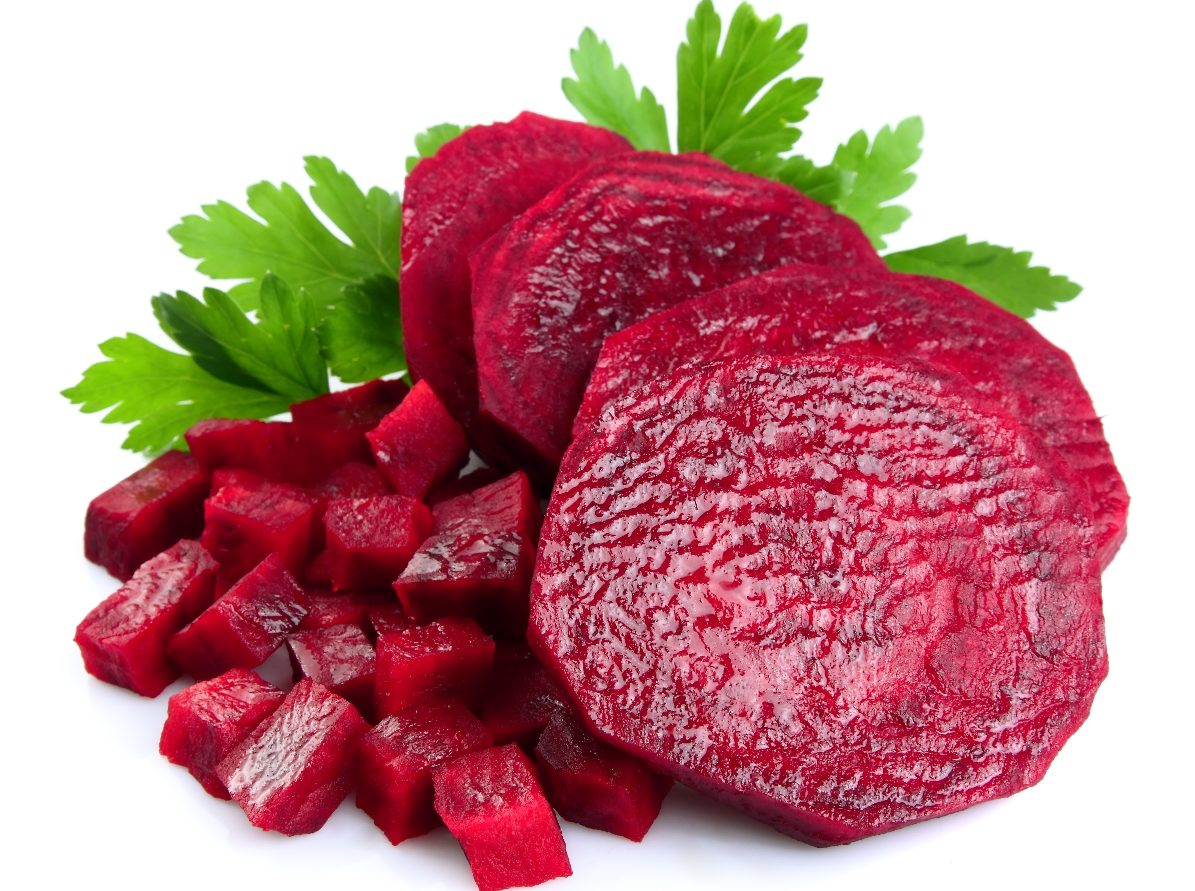
Beet juice in the stools, to some, is “cool,” but to others, is very frightening, as this can masquerade as blood in the stools.
In medicine, “beeturia” refers to the unabsorbed crimson pigment of this vegetable, called betanin, in a person’s urine, not their bowel movements.
If you’re seeing red in your stools three or four days after eating whole beets or beet juice, it might be tempting to start thinking that this is blood.
The betanin comes out in the urine, which comes out with your poops, creating the illusion that your BMs themselves are reddish.
What’s actually happening is that your stools are surrounded by the urine that has the beet pigment in it.
Of course it’s going to get on your bowel movements.
But can this “stool beeturia” still occur five days after you ate the beets or drank beet juice?
Yes — because I myself have had it five days out.
I might also add that I had the beeturia five days in a row, rather than skipping the first four days and then seeing it on day 5.
“In the stool it’s also generally two days, but maybe three or four,” says Jonathan Zinberg, MD, chief of gastroenterology at South Nassau Communities Hospital, Oceanside, NY.
“It is more likely to occur in those who have increased iron absorption, such as those with iron deficiency.
“But remember, not all biologic specimens (i.e., people) follow the rules, so there can be variability.”
For a more in-depth — and reassuring look at this phenomen, read my other article:
Red Stools from Beets vs. from Blood: How to Tell the Difference
 Jonathan Zinberg, MD
 Lorra Garrick has been covering medical, fitness and cybersecurity topics for many years, having written thousands of articles for print magazines and websites, including as a ghostwriter. She’s also a former ACE-certified personal trainer.
Top image: Shutterstock/Volosina
Sharp Sudden Pain in Upper Arm: Non-Cardiac Cause

Find out what can likely cause a sudden sharp pain in the upper arm that’s not related to your heart.
If you’ve been experiencing episodes of a sudden and sharp pain located in your upper arm, this may very possibly be a condition called long head biceps tendonopathy.
This is when the tendon of the long head of the biceps is torn; it may or may not cause pain.
If you have LHB tendonopathy, you may also have a shoulder problem, namely, a rotator cuff tear.
A torn rotator cuff, as well, can cause a sudden sharp pain in the upper arm.
However, these aren’t the only musculoskeletal causes of a sudden sharp pain in the upper arm; hence, a thorough examination, which includes radiographic imaging, will be necessary for precise diagnosis.
The treatment for long head biceps tendonopathy may be surgical or nonsurgical, says Shane Nho, MD, a Chicago-based orthopedic surgeon.
His report appears in the Nov. 2010 Journal of the American Academy of Orthopaedic Surgeons.
Statistics & Trends
-There’s no big difference in patient satisfaction or function between the two main surgical options, which are biceps tenotomy and tenodesis.
-The complication rate from surgery is less than 1 percent.
-Both surgeries can be performed via arthroscopy.
-The JAAOS report urges the pursuit of nonsurgical treatment at first, as this may suffice for ending the upper arm sudden pain and other related issues.
-Younger, active patients should undergo the biceps tenodesis if surgery is warranted.
Conservative Treatment for Long Head Biceps Tendonopathy
Rest, avoiding and/or modifying the offending activity, taking anti-inflammatory drugs, and physical therapy.
Shutterstock/Photographee.eu
If these don’t work, then corticosteroid injections may be done, though these may put a patient at risk for tendon rupture, depending on site of injection.
Besides sudden sharp upper arm pain, long head biceps tendonopathy can also cause the following symptoms:
-Audible snapping or popping in the elbow or shoulder
-Biceps cramping resulting from strenuous use
-Bruising from the middle of upper arm down towards elbow
-Tenderness, pain and/or weakness at elbow or shoulder
-Difficulty turning palm up or down
-Funny bulge above the elbow (from flaccid muscle)
Lorra Garrick has been covering medical, fitness and cybersecurity topics for many years, having written thousands of articles for print magazines and websites, including as a ghostwriter. She’s also a former ACE-certified personal trainer.
.
Top image: ©Lorra Garrick
Source: sciencedaily.com/releases/2010/11/101104101653.htm
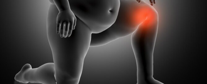
How Effective is Weight Loss for Arthritic Knee Pain?
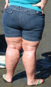
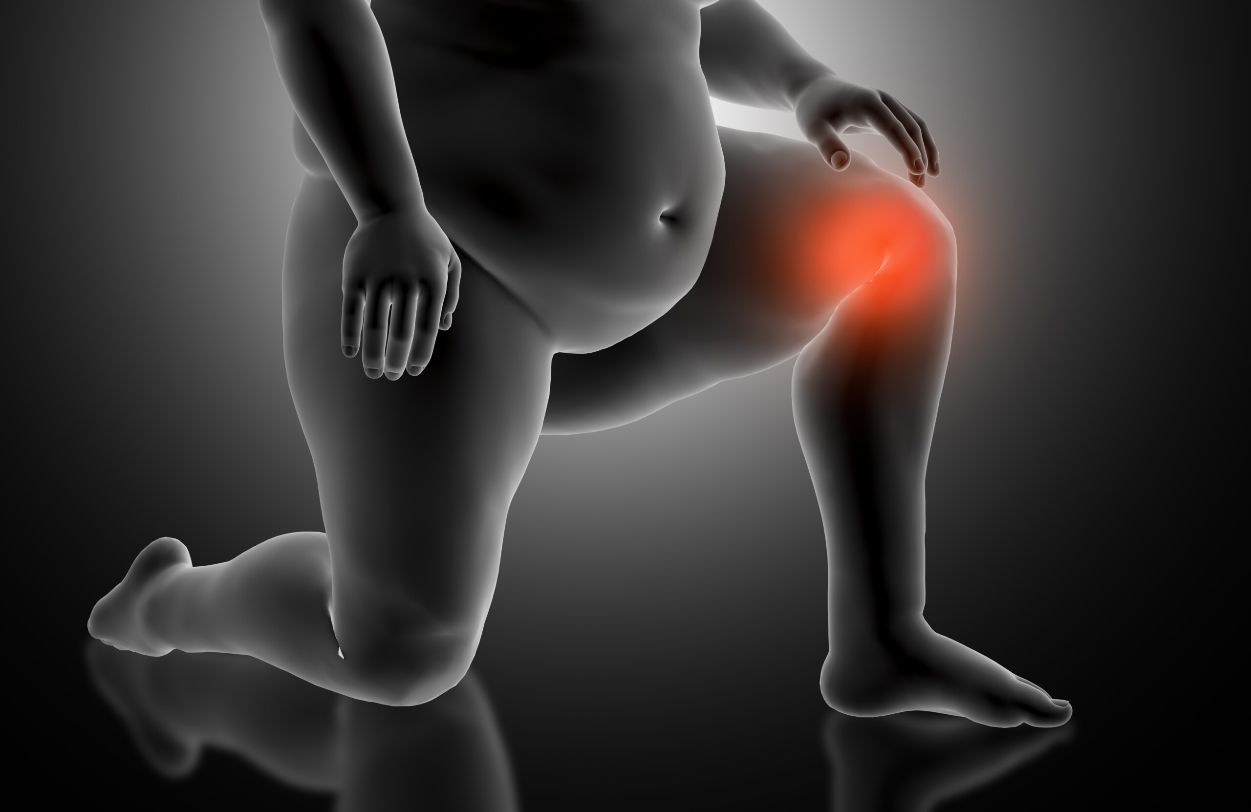
Obesity is one of the leading risk factors for osteoarthritis knee pain; it stands to reason that weight loss will improve the condition.
If you have knee pain, particularly osteoarthritis, and are overweight, then weight loss will probably result in a notable reduction in pain — even though osteoarthritis of the knee can affect medium-built or thin individuals.
Just because thinner people suffer from osteoarthritis of the knee doesn’t mean that obesity isn’t a major risk factor.
The knee is not the most stable joint in the body. Most of one’s body weight is above this joint.
Losing weight will have its virtues for those with painful knees.
Don’t let deluded body-positive people on Instagram trick you into thinking that excess body fat cannot cause issues with quality of life.
- Thin people with knee problems are never told to gain weight for symptom relief.
- Plus size patients are routinely told to drop pounds for symptom relief.
Weight Loss Reduces Knee Pain: Study
Research at the Penn State College of Medicine shows that obese patients who had early-onset osteoarthritis of their knees experienced major improvement in symptoms after an average weight loss of 57 pounds.
Exercising with Knee Pain to Lose Weight
When I was a personal trainer I had overweight clients with knee pain. Losing weight is a must for these men and women.
Aerobic activity can assist with weight loss, but painful arthritic knees will interfere with how rigorous the exercise is.
However, upper body strength training can be done very intensely by overweight people with arthritic knees.
- Lying down bench press and dumbbell press
- Seated chest press with a machine
- Seated overhead dumbbell press or shoulder press with a machine
- Seated cable row
- Seated lat pull-down
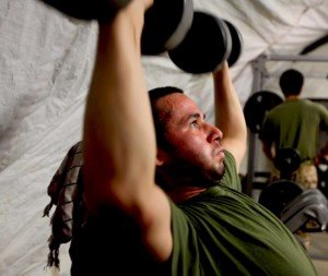
Overhead Dumbbell Press
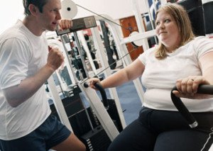
Seated Chest Press
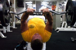
Lying Triceps Extension
As for aerobics, some patients will get a clearance from their doctor for pedaling on a stationary bike or elliptical trainer, plus walking inclines on a treadmill, as long as the arthritis doesn’t cause any pain.
A combination of these aerobics plus weight workouts will facilitate fat loss, which will lead to an improvement in the symptoms of knee arthritis.
Lorra Garrick is a former personal trainer certified by the American Council on Exercise. At Bally Total Fitness she trained clients of all ages for fat loss, muscle building, fitness and improved health.
.
Top image: Shutterstock/Hannamariah
Source: sciencedaily.com/releases/2011/02/110219160015.htm

Rotator Cuff Tear Diagnosis: MR Arthrography vs. Regular MRI

Find out which procedure, the MR arthrography or a standard MRI, is better at detecting a rotator cuff tear.
Diagnosing a rotator cuff tear or other type of tissue tear isn’t always as simple as a patient may think, even if the resulting shoulder pain is classical of this condition.
A study that was done at the Neuroskeletal Imaging in Merritt Island, Florida showed that magnetic resonance (MR) arthrography of the shoulder better identifies a rotator cuff tear, when compared to a regular MRI.
However, not all doctors will inform the patient that the MR arthrography is an option.
When a doctor wanted to figure out what was going on with my mother’s painful shoulder, the only imaging study that was mentioned was the regular MRI.
So if you have a painful shoulder or suspect a torn rotator cuff or some other tear (e.g., labral), be sure to inquire about the MR arthrography, which requires that a contrast dye be injected into the joint.
Says Thomas Magee, MD, the study’s lead author, in the report: “With MR arthrography we were able to see things with a high degree of accuracy in the shoulder.”
Dr. Magee explains that with a non-contrast procedure (3T MRI), the “resolution and picture quality” is high, “except for lesions that are hidden without distension (swelling) of the joint,” he says.
“During MR arthrography, distention of the joint allowed us to uncover lesions that could not be seen on conventional MRI.”
The MR arthrogram provides a “better road map for surgeons,” says Dr. Magee.
If you’ve had only an MRI, and your doctor recommends surgery, WAIT — first insist upon the MR arthrogram for a more accurate assessment.
It may determine you don’t even need surgery.
The report appears in the 2009 American Journal of Roentgenology.
Lorra Garrick is a former personal trainer certified through the American Council on Exercise. At Bally Total Fitness she trained women and men of all ages for fat loss, muscle building, fitness and improved health.
.
Top image: Shutterstock/Andrey_Popov
Source: sciencedaily.com/releases/2009/01/090106154410.htm

Torn Rotator Cuff Pain: Reverse Total Shoulder Replacement
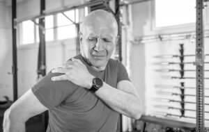
If you have constant pain from a torn rotator cuff, the reverse total shoulder replacement may be a realistic option.
If you have a completely torn rotator cuff and suffer with constant shoulder pain, no amount of physical therapy or cortisone shots will subdue the pain to the extent where you can live a normal life—especially if you also have arthritis in this joint.
The rotator cuff refers to a group of four muscles and their tendons.
They are frequently the subject of some degree of injury, ranging from strain and overuse, to inflammation and impingement syndrome, to a partial or a complete tear.
However, other things can go wrong for which replacement of the shoulder joint is a viable option.
What is the reverse total shoulder replacement and whom is it for?
The shoulder joint is a “ball-and-saucer” structure. In osteoarthritis, there is bone on bone contact, which causes a lot of pain and is “usually associated with joint stiffness,” says Omer llhai, MD, orthopedic surgeon at The Methodist Hospital in Houston.
In osteoarthritis, there is a wearing down of the cartilage between the bones.
The job of this cartilage is to cushion the bones and to provide shock absorption.
When this cartilage deteriorates, the bones begin to rub against each other, leading to pain, stiffness and reduced joint mobility.
The loss of cartilage not only disrupts the joint’s smooth movement but also contributes to the formation of bone spurs and inflammation, exacerbating the discomfort and functional limitations associated with osteoarthritis.
The reverse total shoulder procedure is not designed to remedy a tear in a rotator cuff tendon.
Rather, this surgery is for the replacement of the warn-down saucer part of the joint with a metal ball.
And the worn-out ball part of the arthritic joint gets replaced with a device — this device contains a big plastic cup.
When the saucer is replaced with a metal ball, and the natural “ball” is replaced with the cup, this renders the shoulder joint no longer requiring an intact rotator cuff for motion or stability.
Hence, the patient’s shoulder joint, with proper post-op rehab and physical therapy, should then function normally.
Prevention of a Torn Rotator Cuff
One of the most effective ways to prevent a rotator cuff injury is through strengthening exercises that target the shoulder muscles — in combination with GOOD FORM.
Exercises that enhance the strength and flexibility of the rotator cuff and surrounding muscles can provide better support and stability to the shoulder joint.
Incorporating activities such as resistance training, stretching and range-of-motion exercises into your routine will help maintain shoulder health and prevent injury.
Proper technique and body mechanics during physical activities are crucial for preventing rotator cuff tears.
This includes using correct posture and alignment while lifting weights, participating in sports or performing repetitive tasks.
Reducing the odds of injury can also be achieved if you devote time to warming up your shoulder joints prior to any intense lifting of weights.
Lorra Garrick is a former personal trainer certified by the American Council on Exercise. At Bally Total Fitness she trained clients of all ages for fat loss, muscle building, fitness and improved health.
.
Top image: Shutterstock/Leszek Glasner
Source: sciencedaily.com/releases/2009/03/090325190508.htm

Is Shoulder Surgery Safe for Elderly with Rotator Cuff Pain?

Find out what kind of shoulder surgery is safer for elderly patients suffering with debilitating rotator cuff pain.
An elderly person may be concerned about complications from shoulder surgery.
However, a Rush University Medical Center study shows that for the elderly, arthroscopic surgery is a safe bet, and it works to reduce pain and improve function. Arthroscopy is minimally invasive.
The paper states that for people over the age of 70 who want pain relief, this relief is a fairly predictable outcome of the surgery.
For those of advanced age, pain relief tends to be a higher priority over that of 100 percent restoration of shoulder function.
An example of function is that of being able to retrieve an item from a high shelf, requiring maximum overhead reach of the arms and hands.
Arthroscopy may not restore all the function in some patients, such as those with a large tear in their rotator cuff, but it will go a long way in relieving the pain.
In short, people over age 70 are not too old for shoulder arthroscopic surgery for the treatment of rotator cuff pain.
Tear in the Rotator Cuff
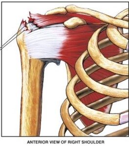
A tear in a rotator cuff tendon. Nucleus Communications, via Wikimedia Commons
“According to a systematic review in 2020, which looked at rotator cuff repair in patients over the age of 70 years, there was a high satisfaction rate with significant improvement in function and pain in this patient population,” says Marc F. Matarazzo, MD, a board certified orthopedic surgeon with Total MD Family Medicine & Urgent Care.
Tears in the rotator cuff occur to about 20 percent of people over age 65.
A surgeon might advise against an operation for elderly patients due to less resilient bone quality and circulation.
But for older people for whom conservative treatments were not effective, arthroscopy is their last hope.
Studies have followed rotator cuff repair patients long-term and found that not only was their pain greatly reduced, but in many cases, their range of motion had improved significantly.
This means being able to raise the surgical arm in front as well as rotate it to the side — motions that were impossible or difficult prior to the procedure.
Another area that the procedure can improve in the elderly is that of strength.
Ninety-four percent of the patients for the Rush University study said they’d have the surgery again if they had to do it over.
Why does arthroscopic shoulder surgery turn out so well for elderly patients?
There are newer surgical techniques, plus the patients’ commitment to post-operative rehab.
However, there will always be patients of elderly age whose rotator cuff tears are determined to be non-repairable, and hence, are not candidates for arthroscopic surgery.
 Dr. Matarazzo specializes in sports medicine and related injuries. He performs minimally invasive and complex reconstructions, and joint replacements, of the shoulder and knee. Dr. Matarazzo is certified in the MAKO robotic-assisted knee replacement system and has 20+ years of orthopedic experience. He has a special interest in cartilage restoration and preservation.
Lorra Garrick is a former personal trainer certified by the American Council on Exercise. At Bally Total Fitness she trained clients of all ages for fat loss, muscle building, fitness and improved health.
.
Top image: Shutterstock/Dmytro Zinkevych
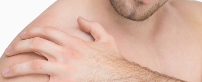
Frozen Shoulder Symptoms vs. Arthritis, Plus Treatment
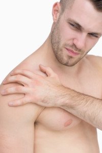
Find out the difference between frozen shoulder and arthritis symptoms, and how to treat frozen shoulder without surgery.
The medical term for frozen shoulder is adhesive capsulitis, and it can be misdiagnosed as arthritis due to the similarity of symptoms.
In frozen shoulder, the tissue around the joint is chronically inflamed; this causes a thickening and tightening in the affected area.
Symptoms of Frozen Shoulder
Significant pain and restrictive movement. These symptoms are also common with other musculoskeletal conditions of the shoulder—such as arthritis.
The right diagnosis may require extensive investigation into one’s medical history.
“Adhesive capsulitis involves idiopathic [unknown cause] inflammation of the shoulder joint capsule, causing pain and decreased shoulder motion,” says Jessalynn Adam, MD, who specializes in primary care sports medicine with OrthoVirginia.
“There are three phases: freezing (the shoulder is painful and stiff), frozen (the shoulder is no longer very painful but motion is significantly restricted), and thawing (shoulder motion gradually returns to normal). This can be quite a prolonged process, up to 26 months.”
Is arthritis sometimes misdiagnosed as frozen shoulder syndrome?
“Perhaps, but age and other health conditions can help to differentiate the two,” says Dr. Adam.
“Frozen shoulder or adhesive capsulitis typically affects middle-aged women between the ages of 40-60.
“It is more common in those with autoimmune conditions such as diabetes, thyroid dysfunction, atherosclerotic/heart disease and Dupuytren’s contracture [a hand condition].”
Painful and Stiff Don’t Always Mean Frozen Shoulder
A report in the 2011 Journal of the American Academy of Orthopaedic Surgeons states that people who have a painful and stiff shoulder are often diagnosed with “frozen shoulder.”
But many things can cause motion loss in a joint.
Details of Frozen Shoulder Symptoms
- Inability to sleep on the affected side due to pain
- Movement so restricted that dressing is difficult, and other things as well like hair care such as shampooing.
- Pain will often decrease once the motion becomes quite restrictive, but pain will bite when the patient makes a sudden movement beyond the confinements of the stiffness.
This is because of microscopic tearing of the scar tissue.
Symptoms of Arthritis
When one tries to move the shoulder, there is often a ratchety or grinding sensation, which is not present with frozen shoulder syndrome.
An X-ray can show arthritis.
Treatment
“Physical therapy focused on capsular stretching, cortisone injected into the shoulder joint to reduce inflammation and pain, rest, time,” says Dr. Adam.
The stretching should be gentle and progressive over a period of weeks, even months sometimes.
“Most cases respond well to these treatments,” says Dr. Adam.
“Refractory cases can be treated with a high volume injection or manipulation of the joint under anesthesia.”
Surgery is a very last resort, but is rarely needed.
 Dr. Adam specializes in the care of athletes and active individuals of all ages, offering prevention, diagnosis and treatment of sports and exercise injuries. Dr. Adam’s care focuses on muscle injuries and biomechanics.
Lorra Garrick has been covering medical, fitness and cybersecurity topics for many years, having written thousands of articles for print magazines and websites, including as a ghostwriter. She’s also a former ACE-certified personal trainer.
.
Top image: Shutterstock/wavebreakmedia
Source: sciencedaily.com/releases/2011/09/110907132100.htm

Lisinopril Can Cause Blackouts and Here’s Why

Yes it’s true that the blood pressure drug Lisinopril can cause you to pass out.
That’s a tough situation to be in: hearing you have high blood pressure but that the medication for this can cause a blackout.
So imagine being told that you need a drug such as Lisinopril to lower your high blood pressure (or increase the “squeezing” ability of your heart) — but that this drug can also cause you to black out as a side effect.
“Yes, Lisinopril can cause blackouts if the person is very sensitive to the medication, meaning their blood pressure drops significantly in response to the medication, and such a precipitous drop can cause one to suddenly black out,” says Dr. Sameer Sayeed, a cardiologist at ColumbiaDoctors of Somers, NY.
It’s also possible that a person can seemingly be compatible with Lisinopril for a very long time — until the state of their body becomes compromised, making them more vulnerable to blacking out or fainting.
An example of that incompatibility would be recent major surgery.
This can make the body more sensitive to Lisinopril, causing a big drop in blood pressure when the patient quickly rises to a standing position — leading to a fainting episode or feeling like the room is “going black.”
How common is fainting from Lisinopril?
“This is not that common with Lisinopril, but can happen especially if the person is dehydrated or not eating or drinking enough, or is already on other BP medications.”
There’s no way to predict ahead of time with certainty whether a given patient will have this particular side effect.
Reducing the Chance of a Blackout
To reduce fainting risk on lisinopril at home, you should stay well-hydrated, first of all. One way to ensure this is to get a very large thermos and fill to the top with water at the start of the day, with the goal of finishing all the water by bedtime — in addition to drinking water with your meals and snacks.
Next, be sure to rise slowly from sitting or lying positions; avoid sudden standing.
Also, limit alcohol (better yet, see if you can quit drinking altogether; it’s not good for the blood vessels), and monitor blood pressure regularly.

Dr. Sayeed performs echocardiograms and stress tests at the Midtown Manhattan and Westchester offices at Columbia Doctors. He is also trained in cardiac CT imaging.
Lorra Garrick has been covering medical, fitness and cybersecurity topics for many years, having written thousands of articles for print magazines and websites, including as a ghostwriter. She’s also a former ACE-certified personal trainer.
.
Top image: Shutterstock/forma82





Can Ejection Fraction Be Increased without Drugs?

What can you do to increase ejection fraction if you don’t want to take medications that might cause fainting?
“There are really no other good ways of increasing ejection fraction other than medications,” says Dr. Sameer Sayeed, a cardiologist at ColumbiaDoctors of Somers, NY.
“Exercise may help to raise ejection fraction, but over the long term is not as proven or effective as ACE inhibitors like Lisinopril or beta blockers such as Carvedilol, which can both cause blackouts.”
If you don’t yet have what would be considered an abnormally low ejection fraction, then what you can do is lead a lifestyle that’s designed to prevent your ejection fraction from getting low enough to necessitate drugs.
Medications do not cure low ejection fraction; there is no cure. Low ejection fraction can lead to congestive heart failure.
Prevention or maximal delaying of this condition comes in the form of avoiding tobacco, engaging in rigorous cardio exercise and strength training, and sticking to an anti-inflammatory diet.

“Aldactone, which is an aldosterone blocking medication, is now also used to help increase ejection fraction after adequate beta blocker and ACE inhibitor doses are already being used,” says Dr. Sayeed.
“It usually does not cause blackouts and may be an option used alone if they cannot tolerate the other two meds.” Aldosterone is a hormone secreted by the adrenal glands.
Dr. Sayeed performs echocardiograms and stress tests at the Midtown Manhattan and Westchester offices at Columbia Doctors. He is also trained in cardiac CT imaging.
Lorra Garrick has been covering medical, fitness and cybersecurity topics for many years, having written thousands of articles for print magazines and websites, including as a ghostwriter. She’s also a former ACE-certified personal trainer.
.