

Thousands of PVCs, Normal Echo: Get EP Testing

A cardiologist says that if you have thousands of PVCs but a normal echo, you should get an EP test for a possible arrhythmia.
If you have PVCs (premature ventricular contractions), you may have developed an understanding along the way that these “skipped heartbeats” are benign as long as your echocardiogram is normal.
“Just because the echo is normal, that doesn’t mean that the person does not have bad coronary artery disease or a PVC focus that is causing all the PVCs,” says Dr. Sameer Sayeed, MD, a cardiologist at ColumbiaDoctors of Somers, NY.
Coronary artery disease can cause these “skipped heartbeats,” and an echocardiogram cannot detect or evaluate plaque buildup in arteries.
Other tests can evaluate for the existence of heart disease or assess heart disease risk: coronary calcium scoring, and the CT angiogram.
“If there are many thousands of PVCs, the patient’s coronary arteries should also be evaluated,” says Dr. Sayeed.
“If they are normal, then EP testing should be performed to determine if there is a PVC focus and if it should be ablated.”
An EP test is an electrophysiological exam of the heart to see if there is a rhythm disorder.
It takes about three hours and is administered by a cardiac electrophysiologist.
A doctor threads thin catheters with electrodes through veins into the heart, records signals and may try to trigger arrhythmias.
This helps diagnose and guide treatments like ablation or pacemaker placement, usually under sedation.
“When there are excessive PVCs, the heart is at risk for an arrhythmia or weakening of the muscle [cardiomyopathy] due to the excessive stimulation by the excess PVCs,” says Dr. Sayeed.
Dr. Sayeed performs echocardiograms and stress tests at the Midtown Manhattan and Westchester offices at Columbia Doctors. He is also trained in cardiac CT imaging.
 Lorra Garrick has been covering medical, fitness and cybersecurity topics for many years, having written thousands of articles for print magazines and websites, including as a ghostwriter. She’s also a former ACE-certified personal trainer.
.
Top image: ©Lorra Garrick

Why a PVC Can Cause Coughing

A cardiologist offers a simple explanation for why PVCs can cause some people to cough.
PVC stands for premature ventricular contraction, and some people can’t help but cough after having one of these episodes.
The coughing is usually just a single or double, and all it takes is just one PVC to trigger this.
“You may cough due to the phenomenon of post-PVC beat where the heart has a stronger contraction after a PVC, and this can irritate the diaphragm or phrenic nerve and cause cough,” explains Dr. Sameer Sayeed, MD, a cardiologist at ColumbiaDoctors of Somers, NY.
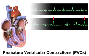
Source: BruceBlaus/CreativeCommons
The phrenic nerves (a pair) come down from the cervical area of the spinal cord and control breathing by innervating the diaphragm.
Both phrenic nerves pass very closely to the heart.
It’s also possible, though less likely than the explanation above, that the cough is triggered by sudden anxiety over feeling a “skipped heartbeat.”
Dr. Sayeed performs echocardiograms and stress tests at the Midtown Manhattan and Westchester offices at Columbia Doctors. He is also trained in cardiac CT imaging.
Lorra Garrick has been covering medical, fitness and cybersecurity topics for many years, having written thousands of articles for print magazines and websites, including as a ghostwriter. She’s also a former ACE-certified personal trainer.
.
Top image: ©Lorra Garrick

Can an Ultrasound Miss a DVT (Deep Vein Thrombosis) ?

It’s very true that an ultrasound can actually miss a DVT, but other tests are more definitive.
Staci Stringer’s DVT was missed by an ultrasound.
In September 2010, she experienced a deep vein thrombosis and pulmonary embolism.
When the DVT developed, Stringer was on birth control, but she also had lupus anticoagulant syndrome and rheumatoid/psoriatic arthritis.
Ultrasound Misses DVT
“It started as a pain in my leg, and after a week I went to the ER and had an ultrasound; they said it was nothing,” says Stringer on her site.
“The following Monday I was admitted to the ER with a PE,” which had caused chest pain.
“After a week or so the PE cut off blood flow to a portion of my lung which caused a pulmonary infarction.”
Another way of saying this is a “heart attack in the lung,” in that oxygen was cut off by the blood clots to the lung tissue, permanently damaging it.
Symptom Detail
“I started to feel the symptoms of a DVT in my right calf,” says Stringer’s account.
“It was swollen, extremely painful, hard to walk. I ignored the symptoms because I figured it was just arthritis pain.”
She continues: “I spoke with my general practitioner after a week and she told me to go to the ER for an ultrasound.”
Ultrasound is a standard diagnostic tool for DVTs.
“The technician thought he saw something but sent me home. I started to get a fever; my calf became so stiff I couldn’t walk.
“I walked up two flights of stairs to my apartment and was so out of breath I was grasping for air for 10 minutes.
“Next day I walked up the stairs to my office and I was so out of breath I fainted. I was admitted to the ER. After the CT scan they found a pulmonary embolism.”
How does an ultrasound miss a DVT?
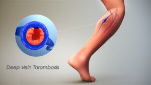
Scientific Animations, Creative Commons/BY-SA/Attribution-ShareAlike 4.0 International
“I regularly order a venous Doppler to rule out DVT,” says Reena Patel, MD, a board certified family medicine physician who treats patients at Garnet Health Urgent Care in NY.
“An ultrasound uses sound waves and compression [of the blood vessel wall] to see blood flow in the veins of an extremity and can find an obstruction.
“There is room for error. Specifically, human error can result in a false negative, so it’s important to see the patient as a whole.”
A probe is used and if it slides off the vessel wall, a false-negative finding can result.
“It’s important in my experience, if suspicion is high and Doppler is negative, to have a backup plan,” says Dr. Patel.
“I may order a D-dimer. This is a sensitive test and can be very helpful to back up your diagnosis and warrant further imaging.
“I consider their history, the current symptoms and calculate their risk using reliable scoring such as the Wells’ score [a numerical value derived from the patient’s answers to numerous questions].”
Additionally, if you believe an ultrasound has missed a DVT, you should ask about a color flow venous duplex scan.
This should be of the vessels close to your pelvis as well as extending down to the foot.
If the D-dimer blood test is positive, the protocol is to have the patient undergo a chest CT scan.
The younger the patient, the more likely that a positive D-dimer means a blood clot, since old age can cause a false-positive with the D-dimer.
What caused Stringer’s DVT?
“The doctors weren’t sure if it was my birth control, my arthritis flare that caused the DVT,” says Stringer, “but I later found out I have lupus anticoagulant syndrome. I’m on warfarin for life.”
 In addition to treating many chronic conditions, Dr. Patel treats urgent conditions that affect every part of the body. Instagram: That_dr_next_door
Lorra Garrick has been covering medical, fitness and cybersecurity topics for many years, having written thousands of articles for print magazines and websites, including as a ghostwriter. She’s also a former ACE-certified personal trainer.
.
Top image: Shutterstock/TANAPAT LEK.JIW

Mild Congestive Heart Failure’s Effect on Exercise

Mild congestive heart failure may not even be noticeable to the person who has it, even when they are exercising.
“Usually if the mild congestive heart failure comes on very gradually and it is only mild diastolic CHF, then it may not be noticeable or only noticeable with very rigorous exertion,” or activity that’s more than usual for the individual, says Dr. Sameer Sayeed, a cardiologist at ColumbiaDoctors of Somers, NY.
So if you’ve always gone out every morning to leisurely walk your dog for half a mile, and all along your diastolic heart function is getting a little worse such that it’s in the range of mild heart failure — you probably won’t be suddenly panting at the end of that half mile.
Diastolic CHF: The heart is stiff, not relaxing properly. It does not fill up with the right amount of blood to be pumped out.
Thus, less blood is pumped out, meaning less oxygen throughout the body.
“If it is more severe diastolic CHF, then it may be a more acute and rapid decline,” says Dr. Sayeed.
“If it is systolic CHF and the ejection fraction [pumping force] goes below 40 percent, then the symptoms may come on more rapidly and with minimal exertion like [walking] a block or two or one flight of stairs.”
Systolic CHF: decreased pumping function. The heart’s “squeeze” is not optimal.
Dr. Sayeed explains, “The fatigue is usually most noticeable with exertion, particularly stairs and inclines, and the patient may suddenly report not being able to go up stairs or a hill or do competitive sports, etc.
“Symptoms won’t be so apparent at rest or with things like golf or easy activities where it may go unnoticed.”
If you’ve been working out all along, it’s smart to keep a record of your key workloads, which may be measured in amount of resistance, distance, speed or time.
As we age, these values will gradually decrease, e.g., your fastest one-minute run on a treadmill may be 12 mph at age 50, but 11.5 mph at age 55. This is normal decline.
If a performance deficit, however, is suspiciously rapid with no explanation (such as recent injury), then this warrants a medical evaluation.
Congestive (chronic) heart failure can be one of numerous causes of an inexplicable deficit in aerobic stamina or strength.
Dr. Sayeed performs echocardiograms and stress tests at the Midtown Manhattan and Westchester offices at Columbia Doctors. He is also trained in cardiac CT imaging.
Lorra Garrick has been covering medical, fitness and cybersecurity topics for many years, having written thousands of articles for print magazines and websites, including as a ghostwriter. She’s also a former ACE-certified personal trainer.
Can Congestive Heart Failure Cause Only Edema?
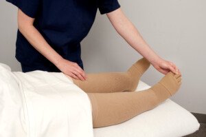
Edema refers to fluid retention such as that in the lower legs that causes swelling, but can this be the ONLY symptom of congestive heart failure?
Can a person feel and function just fine otherwise, despite the puffy lower legs and ankles from the congestive heart failure’s edema?
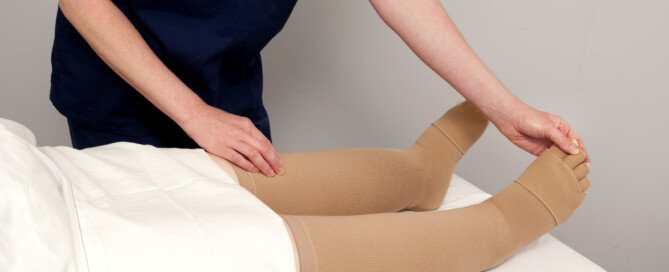
Congestive heart failure can cause numerous symptoms, including the fluid retention of edema.
Sometimes, this backed-up fluid goes higher than knee level, affecting the upper legs and even the groin area.
“Congestive heart failure can just cause edema and no other symptoms if it is due to purely right [heart] sided failure,” says Dr. Sameer Sayeed, a cardiologist at ColumbiaDoctors of Somers, NY.
“But this is rarer, as right sided failure is usually caused by left heart failure which would cause dyspnea.”
The “failure” refers to the heart’s right or left ventricles failing to perform efficiently. And dyspnea means shortness of breath.
In heart failure, the pumping action of this organ is either inadequate, and/or the heart’s chambers don’t refill with as much blood as they should prior to each beat.
The end result is an insufficient supply of oxygenated blood circulating throughout the body.
So what this all means is that many more times than not, a patient will have, in addition to edema from the congestive heart failure, a problem with shortness of breath or getting out of breath fairly easily.
For example, activities that historically did not leave the patient breathing hard, now leave him or her winded, such as walking up a flight of stairs, walking quickly on an inclined parking lot, doing housework or lightly playing with the grandkids in the backyard.
They will fatigue more quickly from day-to-day activities that had never tired them out before.
When the excess fluid is in the groin area, it can impose upon the bladder, reducing urine output. This fluid would be visible on a CT scan.
Other Symptoms of Heart Failure
In addition to edema and shortness of breath, CHF can cause nausea, appetite suppression, a cough with white or pinkish phlegm, and chest pain.
Consult with your cardiologist if you have any concerning symptoms including new-onset ankle or lower leg swelling in BOTH legs, which could signal congestive heart failure.
If you have new-onset swelling in only one leg, particularly the lower leg, this could be a deep vein thrombosis.
This needs immediate attention, as this blood clot could detach and very quickly travel to the lungs and be life threatening.
Dr. Sayeed performs echocardiograms and stress tests at the Midtown Manhattan and Westchester offices at Columbia Doctors. He is also trained in cardiac CT imaging.
Lorra Garrick has been covering medical, fitness and cybersecurity topics for many years, having written thousands of articles for print magazines and websites, including as a ghostwriter. She’s also a former ACE-certified personal trainer.
.
Top image: Shutterstock/futurewalk
Source: mayoclinic.com/health/heart-failure/DS00061
Fast Rotator Cuff Tendonitis Pain Treatment without Surgery

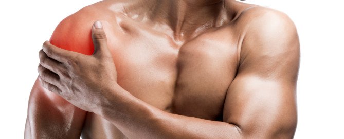
The procedure is minimally invasive, nonsurgical and provides immediate pain relief from rotator cuff tendonitis.
A minimally invasive procedure to treat tendonitis in the rotator cuff provides immediate symptom relief, according to a study.
For some patients, their rotator cuff can accumulate calcium residue and be resistant to physical therapy.
“Calcific tendinopathy often resolves on its own but can become chronic,” says Jessalynn Adam, MD, who specializes in primary care sports medicine with OrthoVirginia.
“Often, a combination of physical therapy and shoulder injection with/without percutaneous needle tenotomy is sufficient treatment.
“The U.S.-guided procedure is effective but is uncomfortable. Patients are often sore following the procedure for a couple of days as well. However, there are good results.
“A study demonstrated that at one year, patients who underwent needling had more improvement than simple cortisone injection alone.
“Surgery is uncommon for this condition and only indicated for patients with refractory symptoms for six months or more (about 10% of cases).”
Another study found that ultrasound-guided nonsurgical therapy significantly reduces pain from calcific tendonitis of the rotator cuff and restores lasting mobility after treatment.
The treatment resulted in a single and inexpensive approach that was effective, says the study’s author, Luca M. Sconfienza, MD.
In severe cases, patients may require shockwave treatment or open surgery to remove the calcium.
Open surgery requires a hospital stay and rehabilitation and, on rare occasions, may result in major complications, such as tendon rupture.
How the Procedure Is Done
For the 20-minute procedure, the shoulder is anesthetized and, with ultrasound guidance, a radiologist injects a saline solution into the rotator cuff to wash the area and break up the calcium.
A second needle is used to aspirate, or withdraw, the calcium residue. Recovery time is about an hour.
Complete absence of the pain, following only a single procedure, is not guaranteed, but may be well worth it for chronic sufferers.
Symptoms of Rotator Cuff Tendonitis
• Pain in the upper side of the arm when lifting the arm straight out at one’s side
• Same pain when reaching overhead, especially with straight arms.
• Same pain when slipping the arm through the sleeve of a coat or jacket.
 Dr. Adam specializes in the care of athletes and active individuals of all ages, offering prevention, diagnosis and treatment of sports and exercise injuries. Dr. Adam’s care focuses on muscle injuries and biomechanics.
 Lorra Garrick has been covering medical, fitness and cybersecurity topics for many years, having written thousands of articles for print magazines and websites, including as a ghostwriter. She’s also a former ACE-certified personal trainer.
.
Top image: Shutterstock/xmee
Source: sciencedaily.com/releases/2009/06/090630074949.htm

Parkinson’s: What Cardio Exercise Intensity Works Best?

Find out what a study says about the best cardio intensity level for Parkinson’s patients.
Will those with Parkinson’s disease benefit more from low intensity cardio exercise or higher?
Researchers at the University of Maryland School of Medicine and the Baltimore VA Medical Center wanted to find out what would be a preferable exercise for those with this neurological disorder.
Study Results
The low intensity group experienced the greatest degree of consistent improvement in gait and mobility.
For the study, subjects with Parkinson’s disease walked on a treadmill and also did resistance and stretching exercise.
There were three groups:
1) 50-minute treadmill walk for low intensity, 2) 30-minute incline walk for high intensity, 3) resistance exercise for the legs.
All Parkinson’s groups exercised for three months, three times a week.
“These results show that exercise in people with Parkinson’s disease can make a difference in their function,” explains Lisa Shulman, MD, the lead study author, professor of neurology at the University of Maryland School of Medicine.
In the report she adds, “Exercise may, in fact, delay disability and help to preserve independence.”
People with Parkinson’s should strongly consider low intensity cardio workouts.
Walking on a Treadmill for Parkinson’s Disease
Based on this study, it appears that treadmill walking will produce the greatest benefits for ambulation, when compared to using other kinds of cardio equipment.
After all, to get from point A to point B, we usually walk rather than pedal or step upward.
Walking is the most natural form of human movement, so the treadmill is the obvious choice for improving ambulation.
As far as cardiovascular benefits, any cardio equipment will suffice.
Getting Started on a Treadmill
Those with Parkinson’s disease should begin slowly on the treadmill and work on avoiding the habit of continuous holding on, as this will reproduce a gait pattern similar to that of using a walker. The image below demonstrates this.
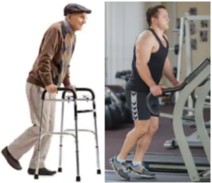
Continuous holding on can also lead to repetitive stress injuries in the hips, plus “mold” posture into an incorrect form.
As your balance improves, gradually increase speed.
Again, the key is starting out at a slow-enough speed rather than a speed that makes you feel too off-balance to let go.
When I was a personal trainer, I noted that whenever someone told me they feared falling off a treadmill unless they held on, they had the speed at least 3.5 mph and often had a high incline.
If you must start out at only 1.5 mph, or even just one mph, at zero incline, then do so.
Then from there, gradually increase your speed over time, but never too much that you feel that you’ll lose your balance.
Holding on momentarily for balance checks is encouraged, but again, do not get into the habit of CONTINUOUSLY holding on — and this assumes that you can already walk in day-to-day living without a walker or cane.
Lorra Garrick is a former personal trainer certified by the American Council on Exercise. At Bally Total Fitness she trained clients of all ages for fat loss, muscle building, fitness and improved health.
Top image: Shutterstock/Kristiana Gankevych
Source of Parkinson’s study summary: sciencedaily.com/releases/2011/04/110412162404.htm
Blood Clots (DVT): Q & A with MD about this Silent Assassin
Dr. Darren Klass answers questions about deep vein thrombosis, a potentially fatal type of blood clot that can strike anybody, any fitness level, any age.
Darren Klass, MD, is Clinical Instructor of Interventional Radiology at the University of British Columbia and the Vancouver Coastal Health Association.
In layman’s terms, what is DVT?
DVT is deep vein thrombosis; this term generally refers to the development of a clot in the deep veins in the legs and pelvis.
The deep veins in the legs run between muscle compartments and cannot be seen without medical imaging.
How is it caused?
DVT is caused by blood within moving veins clotting. The clot is cause by three factors:
- Slow flow in the vein
- Abnormal clotting factors in the blood which cause it to clot more than normal
- Damage to the lining of the vein
Are there ways to prevent?
There is no way of completely preventing blood clots without medication, which in itself has risks. Ways to decrease the risks are:
- Keep well-hydrated; dehydration thickens the blood.
- Avoid sitting in the same position for long periods of time (i.e., long plane flights).
- Avoid tight constricting clothing at the hip, which may squash the vein in the groin and slow flow.
How can I be on the lookout for a clot?
If one part of your leg (the calve, thigh or both) suddenly becomes swollen and painful, or suddenly veins which were not visible under your skin become more prominent, you should consult your family doctor.
What are the risks if it’s not caught?
DVT may cause little or no long term complications if treated early. If left untreated, the risks are the following:
- Piece of the clot may break off and travel to the lung causing a pulmonary embolism.
- The clot may extend into the main vein in the abdomen (termed the IVC) and stop the flow of blood from the pelvis and opposite leg, which may cause complications.
- If the clot involves the veins in the pelvis, this may require treatment in a hospital to break up the clot (thrombolysis performed by an interventional radiologist). The aim of the treatment is to clear as much clot from the veins as possible and allow healing of the vein.
If the vein is left untreated and the clot is absorbed by the body, the vein is often left scarred.
The scarring may damage valves in the vein, which can lead to a chronic swollen leg, which is difficult to treat and may cause significant limitation in everyday activity. This condition is called post-phlebitic syndrome.
What are the options for treatment of DVT?
DVT can be treated in the following ways:
Medication to thin the blood and decrease the risk of the clot extending.
However, the clot remains in the vein until the body reabsorbs it and the vein is often left scarred.
The clot can be removed by a minimally invasive procedure performed by an interventional radiologist.
The procedure involves using special medication and advanced equipment to break up the clot and remove it from the vein and restore flow immediately.
The procedure is done through a tiny hole made in the vein behind the knee and a very small cut in the skin about 3 mm in length.
 Dr. Klass specializes in interventional oncology, aortic intervention, PVD, venous disease and venous access. He has been inducted into The Leading Physicians of the World, published by the International Association of HealthCare Professionals.
Lorra Garrick has been covering medical, fitness and cybersecurity topics for many years, having written thousands of articles for print magazines and websites, including as a ghostwriter. She’s also a former ACE-certified personal trainer.

Cause of Heavy Bleeding after Missed Period

The cause of heavy bleeding in menstruation that follows missed periods is quite logical.
Many women wonder and are even worried about the cause of heavy flowing in a period after menstruation has been missed once or a few times.
“The cycle consists of estrogen stimulating the lining of the uterus (endometrium) and ‘building up’ the lining,” begins Dr. Marlan Schwartz, MD, FACOG, robotic surgeon with Lifeline Medical Associates.
“If this is not shed, and there are multiple reasons that could cause this, it will likely be kept being stimulated by more estrogen, thus causing the lining to be even thicker.
“If one then has a period or loss of this lining, as it has been made thicker (six or seven weeks of stimulation versus three weeks), there is more to shed.”
 Dr. Schwartz is the past Chairman of the Department of Obstetrics and Gynecology at Robert Wood Johnson University Hospital-Somerset.
Lorra Garrick has been covering medical, fitness and cybersecurity topics for many years, having written thousands of articles for print magazines and websites, including as a ghostwriter. She’s also a former ACE-certified personal trainer.





Are There Reasons a Colonoscopy Can Miss Colon Cancer?
You know a colonoscopy can miss colon cancer, right? So find out exactly why, and what questions to ask your doctor.
Though a colonoscopy has saved the lives of many people by detecting precancerous polyps, it can also miss actual colon cancer.
“Endoscopy is not perfect and that’s tough fact to deal with as a gastroenterologist,” says Whitney Jones, MD, a national expert and frequent speaker on early-age onset colon cancer prevention, and Founder, Colon Cancer Prevention Project.
For average-risk people, colonoscopies should begin at age 45 — according to the U.S. Preventive Services Task Force — and be done every 10 years after.
“The 10 year interval as we all know is a negotiated time frame, not a date set in stone from God,” says Dr. Jones. “It’s the best estimate.”
The truth is, a colonoscopy can provide “incomplete protection,” adds Dr. Jones, which “usually lies with three factors.”
Three reasons a colonoscopy can miss colon cancer
#1. “Operator characteristics: Not all docs who do endoscopy do the procedure equally well,” says Dr. Jones.
“Many are benchmarking and working on continuous improvement, i.e., tracking fecal intubation rates, withdrawal times, adenoma [cancer] detection rates, of using split dose preps which better clean and prepare the right [sided] colon.
“These are questions any person having a colonoscopy should ask their physician.”
The second reason a colonoscopy can miss colon cancer is “anatomic considerations,” says Dr. Jones.
“It is impossible to see every square cm of the colon with any modality. There is a miss rate of up to 10% for 1cm adenomas, even by experts.”
Reason #3 for why a colonoscopy can miss colon cancer: “Biological issues: Right colon adenomas are much more likely to have an evolving pathology called serrated adenomas; these polyps evolve into cancer through a different pathway called hyper- methylation. They are not your mother’s colon polyps/cancers.
“They have several differences including a more flat nature, making them harder to identify, be obscured with mucus or stool, progress to cancer in a shorter time frame and be located obviously in the right colon, which is harder to reach.”
Dr. Jones advises high-risk people to request more frequent screening and to consider asking their doctor for a stool test between colonoscopies.
 Dr. Jones’ practice interests include prevention and treatment of colon cancers, pancreatic disease and biliary disease. He has authored numerous scientific articles, reviews and abstracts and presented at a variety of national and international scientific meetings. Colon Cancer Prevention Project
Lorra Garrick has been covering medical, fitness and cybersecurity topics for many years, having written thousands of articles for print magazines and websites, including as a ghostwriter. She’s also a former ACE-certified personal trainer.
.