

Melanoma Risk with Fair Skin, Brown Eyes vs. Blue Eyes

Find out if there’s a difference in melanoma risk in someone with fair skin and brown eyes vs. fair skin and blue.
Lists of melanoma risk factors often include “fair/light skin and blue eyes.” Sometimes, the list says, “blue or green eyes.”
Melanoma risk lists always name fair or light skin, or Northern European descent—which implies very light tone.
This all begs the question: Is melanoma risk higher in a person with light skin and blue eyes, when compared to an individual with light tone and brown eyes?
“I don’t believe there was ever a statistical difference found (or even investigated) regarding the color of eyes in a person who is fair skinned, as related to their risk of developing melanoma,” says Judith Hellman, MD, Associate Clinical Professor of Dermatology, Mt. Sinai Hospital, NY, who has a private practice in Manhattan.
“It is clear that the lighter the color of a person’s skin, the less protection it has against sun damage,” continues Dr. Hellman.
“A lighter color of hair and/or blue eyes probably just signal that the propensity of that person’s body is to produce less melanin, which is what provides the dark color of the skin, and which is what protects the skin from UV radiation.”
In other words, the more melanin in a person’s skin, the darker the tone will be, and of course, the more likely they will have brown eyes.
However, the color of brown may have nothing to do with the risk of this deadly cancer, and instead, brown is merely a marker for how much melanin a person’s body produces.
Keep in mind that many light or fair people have brown eyes, including Asians and Northern Europeans.
However, a person with dark pigment who has green eyes has a lower risk of melanoma (when basing risk on skin tone) than the Asian or Northern European with very light skin who has dark brown eyes.

Light skin and brown eyes vs. light with blue eyes, and the melanoma risk.
Dr. Hellman explains, “So while the risk may be a tad higher, I don’t believe it’s something that a study would demonstrate.
“Common sense dictates that the lighter the skin, hair and eye color, the more a person should be aware of the risk of melanoma and be vigilant about getting regular skin exams.”
 Dr. Hellman practices medical dermatology and also specializes in laser surgery, anti-aging skin treatments and dermatologic surgery.
 Lorra Garrick has been covering medical, fitness and cybersecurity topics for many years, having written thousands of articles for print magazines and websites, including as a ghostwriter. She’s also a former ACE-certified personal trainer.
.
Top image: ©Lorra Garrick
Source: BigAppleSkin.com

Benign Cause of Brief Stabbing Pains in Breast

Find out a most unlikely, but harmless, cause of sudden-onset, brief stabbing pains in a breast; and it’s not a tight bra.
- Have you developed sudden-onset, stabbing, sharp or piercing pains in a breast?
- And do these stabbing pains in your breast last maybe one to 10 seconds?
- Do they come on with seemingly no provocation? No particular position (walking, jogging, bending over, stretching, reaching) brings them on?
Ask yourself this question: Has your arm been pressing up against the breast lately? This is what happened to me. I developed very brief shooting, severe pains in my breast.
I immediately suspected what I call “compression.” This is when my arm “compresses” the breast by leaning or pressing into it.
In the past, this would happen when I slept overnight on my parents’ couch when staying over. The couch isn’t wide enough for me to lie on my back with both arms straight out at my sides on the couch.
The right arm is forced by the couch’s back support to go up on my tummy. When this happens, the upper portion of the arm presses into the outer quadrant of my breast.
Later on in the day I’d notice on and off, or fleeting sensations or aches in that area of the breast. But they were never stabbing or sharp kinds of pain.
However, when I took to massaging my elbow a certain way (I have golfer’s elbow), I began getting shooting piercing jolts in that outer quadrant, as well as middling through vertically.
I’d lean back into my sofa. The arm of my massaging hand was draped over the breast and pressing into it, in order for my fingers to get a firm grasp on the opposite elbow area; the arm being massaged would rest in the sofa.
During these sessions, there was no breast discomfort. But soon after I’d begin feeling some discomfort coming and going.
One day it escalated like mad and took my breath away. I’m talking JOLTING, BOLTS of pain that lasted one to 10 seconds.
I immediately backed off on the particular massaging position, and predictably, the pain began dissipating. But then I’d be back to the positioning, and the problem kicked up again.
Finally, I decided to abandon the position altogether and massage the elbow another way.
The breast pain completely disappeared, but it took about 10 days, dwindling with each day as far as frequency and severity.
When I went in for my routine physical, I mentioned this to the doctor. She wasn’t the least bit concerned.
She did NOT say anything like, “Gee, we’d better get the breast imaged right away,” or, “I don’t like the way that sounds,” or, “That’s likely a sign of something bad.”
No sir. Her demeanor was as though I were describing pain from a first-degree burn from the stove. I asked her what about the “compression” could cause stabbing pain in the first place?
The doctor wasn’t able to offer an explanation as far as the dynamics, other than to say it could have involved nerves and sensitive breast tissue.
She didn’t think it involved ligaments, but DID say it could have involved the chest muscle. She likened the situation to the pain some women get when wearing an underwire bra.
The sharp, stabbing pain in my breast has disappeared, coinciding with cessation of the compression positioning.
Take a good look at your position habits and see if at any time your arm is pressing into your breast.
The resulting pain can be delayed by hours. If the problem persists despite making a correction to your positioning, see your doctor.
Lorra Garrick has been covering medical, fitness and cybersecurity topics for many years, having written thousands of articles for print magazines and websites, including as a ghostwriter. She’s also a former ACE-certified personal trainer.
.
Top image: Freepik/jcomp

Bilateral Ankle Edema: Causes when Tests Are Normal
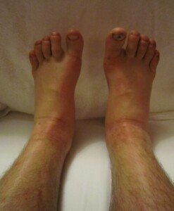
A doctor explains what can cause swelling (edema) in both ankles when your heart, liver, kidney and other key tests are normal and you’re not obese.
The following can cause swelling (edema) in both (bilateral) ankles: heart, liver and kidney failure; low thyroid; celiac disease; kidney stones; PMS; excessive sitting or standing; hot weather; varicose veins; high salt (sodium) intake; old age.
Tests for heart, liver, kidney and thyroid function, as well as a blood test for celiac disease, can easily be taken. What if they’re normal?
Does that mean there can’t be other causes of bilateral ankle edema?
“Ankle swelling in general is common, and when painless can be caused by such simple things as gravity, such as when one is on their feet for long time,” says Jacob Teitelbaum, MD, medical director of the Fibromyalgia and Fatigue Centers nationally, and author of “The Fatigue and Fibromyalgia Solution.”
“Low thyroid will also be a common cause, despite normal thyroid tests.” So if your thyroid test was normal and there seems to be no other cause to the swelling in both your ankles, what should you do?
“Relying on thyroid testing misses over 50% of people who need and would benefit from thyroid hormone,” continues Dr. Teitelbaum.
“In this situation, however, there will also be fluid in the fingers, which would not be especially noticeable except for causing rings to be tight.
“The key discriminant factor also is whether the swelling in the ankles is gone in the morning after one has been lying down for an extended period (i.e., with gravity draining their legs),” says Dr. Teitelbaum.
“If this is the case, there is a greater likelihood of the issue being benign. If the swelling is persisting even through the night, or swelling is very marked even just during the day, I would consider a CAT scan of the pelvis and abdomen looking for evidence of venous obstruction – especially from tumors.”
Keep in mind, though, that bilateral ankle swelling is not a more common symptom of cancer, and it’s not even listed by Mayoclinic.com as a symptom of liver, kidney, lung, colon, stomach (esophageal), ovarian or cervical cancer.
However, Mayoclinic.com lists “swelling in the legs” as a symptom of prostate cancer, which also can yield many other symptoms.
A tumor that obstructs lymphatic flow can cause edema in the ankles, but it’s very unlikely that this would be bilateral.
Periomenopause is associated with bilateral ankle edema. Of course, pregnancy can cause this. But what about menopause?

Shutterstock/fizkes
Menopause and the timeline soon after, during which women can still experience annoying symptoms related to menopause, can cause fluid retention in the body.
Edema in both ankles has been associated with menopause.
The key is determining when you think you began developing the edema, and if this coincides with the timeline right before you began menopause, or during menopause.
If “all the tests” have come back normal regarding the cause of your bilateral ankle edema, consider that pulmonary hypertension can cause this symptom.
However, this condition yields other troublesome symptoms such as dizziness, chest pain and fatigue.
Venous insufficiency can cause swelling in both ankles, but this is usually accompanied by leg pain as well.
Sometimes, the cause of bilateral ankle edema is never found.
Dr. Teitelbaum is a board certified internist and nationally known expert in the fields of fibromyalgia, chronic fatigue syndrome, sleep and pain.
 Lorra Garrick has been covering medical, fitness and cybersecurity topics for many years, having written thousands of articles for print magazines and websites, including as a ghostwriter. She’s also a former ACE-certified personal trainer.
.
Top image: James Heilman, MD, CreativeCommons
.
Sources:
mayoclinic.com/health/prostate-cancer/DS00043/DSECTION=symptoms
mayoclinic.com/health/pulmonary-hypertension/DS00430/DSECTION=symptoms

Sudden Brief Extreme Dizziness in Younger Adults: Causes, Solutions
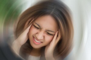
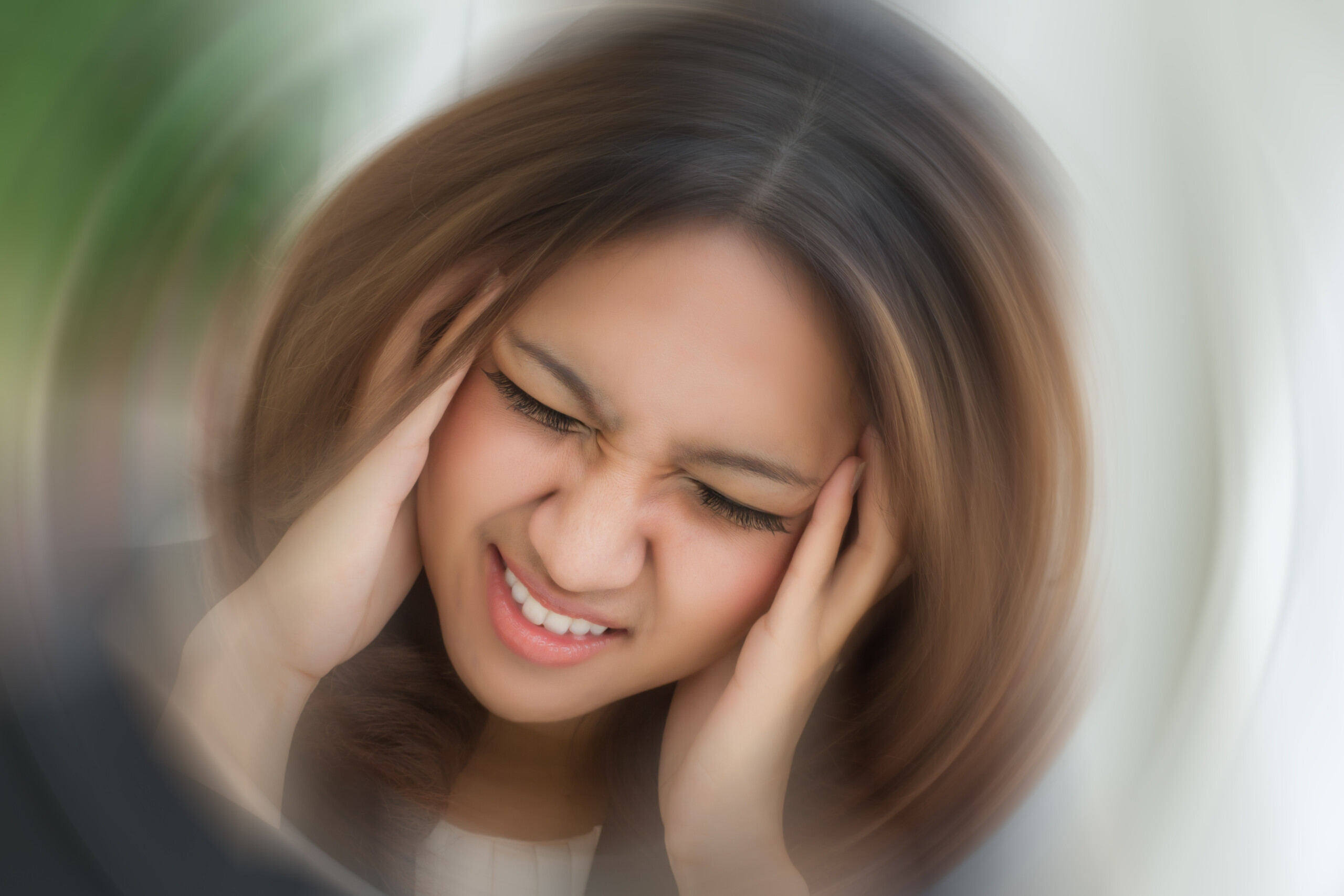
A doctor discusses possible causes of sudden, extreme and brief dizziness in younger people. And solutions.
There are MANY causes of sudden, brief episodes of severe dizziness in people who do not have any conditions that can cause this — that is, conditions that are strongly associated with elderly age.
“Most common ones include abnormal heart rhythms, neck, muscle spasms, ear infections, drops in blood sugar and mini-strokes,” says Jacob Teitelbaum, MD, medical director of the Fibromyalgia and Fatigue Centers nationally, and author of “The Fatigue and Fibromyalgia Solution.”
A mini-stroke is also known as a transient ischemic attack. Though elderly age is a big risk factor, middle-aged people and even younger adults can experience a TIA, though in younger adults, this is rare.
A TIA can cause many symptoms, and though a sudden and brief episode of severe dizziness is one of them, it’s not one of the more common symptoms of a mini-stroke.
In fact, if you’re middle aged and especially younger, and you’ve been experiencing sudden-onset dizziness with no other symptoms, this is unlikely to be a TIA, but a checkup with a neurologist would be a smart idea.
Middle Ear Problem As Cause of Sudden-Onset Severe Dizziness
Dr. Teitelbaum says, “A key discriminating factor? Do they have vertigo where it feels like they or the room are actually spinning around in a circle?
“If yes, this points to a middle ear problem. Also, do they pass out without warning? This suggests an abnormal heart rhythm.”
Side effects from medications (especially narcotics), and pregnancy, can cause a bout of sudden intense dizziness.
There are different kinds of “dizziness.” One such type is called orthostatic hypotension.
Do you get your symptom only right after you suddenly stand after having been seated for a while?
If so, rise slowly from your seat and when you’re halfway up, pause for 15 seconds, then resume rising to an erect position slowly.
This will prevent a sudden drop in blood pressure, which can cause acute dizziness and even blacking out in some individuals.
BPPV
Benign paroxysmal positional vertigo (BPPV) is a common inner ear disorder that triggers sudden, short (or long) episodes of dizziness or a spinning sensation.
It can certainly occur in young adults and people of all ages.
It occurs when tiny calcium crystals, called otoconia, become dislodged from their usual position in the utricle of the inner ear and move into the semicircular canals of the inner ear.
These canals help detect head motion. When the crystals shift with certain head movements, they send false signals to the brain, conflicting with normal balance input.
This mismatch can cause abrupt vertigo lasting briefly in some cases, often brought on by rolling or sitting up in bed, looking up or sideways, or bending over.
Dr. Teitelbaum is a board certified internist and nationally known expert in the fields of fibromyalgia, chronic fatigue syndrome, sleep and pain.
Lorra Garrick has been covering medical, fitness and cybersecurity topics for many years, having written thousands of articles for print magazines and websites, including as a ghostwriter. She’s also a former ACE-certified personal trainer.
.
Top image: Shutterstock/9nong

Are You Rude Due to Your Hearing Loss?

If you have hearing loss, here’s why people get irritated when you ask them to repeat or why they say “Never mind, it’s not important.”
Many people with hearing loss, impairment, who are hard of hearing, etc., report that many individuals get irritated or become rude when asked to repeat something or speak louder.
I’ve seen this situation come up in advice columns: A hearing impaired individual writes in to complain that, so often, people become irate and impatient when that individual asks them to talk slower, louder or to repeat what they just said.
One such woman with hearing loss said it was hurtful when people would respond, “Never mind, it’s not important,” when she asked them to repeat their statement or question.
To all such men and women with hearing loss who have experienced rudeness or exasperation from people, I have a question for you:
HOW do you ask people to speak louder, slower or repeat things?
Take a good look in the mirror and honestly answer this very fair question.
I know people with hearing loss. I can’t begin to tell you how many times I’ve witnessed one of them, a woman I’ll call “Linda,” respond rudely to a speaker when she couldn’t understand them!
She’ll typically deliver a curt “What?!” with an anguished face when she can’t understand what was just spoken to her.
This makes the speaker think that Linda blames THEM for why she didn’t hear them clearly!
Think about it: You’re talking to someone (and, in this hypothetical scenario, you can hear perfectly). That person reacts with an exasperated “What?!” and looks miffed.
How would this make you feel? Would you want to kindly, patiently repeat yourself?
Or would you be totally put off and decide to end the interaction with a “Never mind, it’s not important”?
People do not want to feel as though the hard of hearing listener blames them for why they can’t understand their speech.
Speakers who are made to feel that the listener blames them for “mumbling” or talking unclearly will find this very offensive and rude.
Granted, some people DO mumble and talk unclearly, but let’s face it: Most people do not have this problem.
I know I don’t. In fact, I’ve been told I sometimes talk too loud. As a person who communicates (writing) for a living, I place a high premium on the importance of clear speech. I don’t drink or take drugs; my speech is very clear.
One time I had a phone job. I called someone and began speaking. The cranky man interrupted with a blunt, angry “Speak up! I can’t hear you!”
Well, I didn’t speak up; I HUNG up. I’m sure he thought I was rude!
Had he interrupted with a kindly “Excuse me, but can you repeat that and talk a bit slower and more careful? I’m having trouble hearing you,” I would have kindly obliged.
I know a relative with hearing loss who wears hearing aids, but he often gets irate with me if he doesn’t understand me. He accuses me of “running my words together.”
If you have difficulty with hearing, have you ever accused the speaker of “mumbling” or talking too fast?
This is VERY insulting and provides NO incentive for the speaker to be kind and patient with you.
If you think only children mumble, realize that kids’ voices have higher frequencies, and that’s why you have more difficulty deciphering kids’ speech.
Linda thinks nowadays, everyone on the phone who leaves messages “can’t talk clearly.”
The man with hearing aids I mentioned believes that “today’s movies have poor sound quality,” that the actors don’t speak loud enough and mumble.
He also thinks that “young people don’t know how to leave clear phone messages.”
You’re pretty much guaranteed a kind, thoughtful, patient response from the speaker to repeat or talk slower if you do your part:
Be gracious when asking someone to repeat their question or statement, without implying that they can’t talk clearly.
Don’t be curt just because the speaker is a teenager or child, either. And remember, children emit higher frequencies.

Shutterstock/locrifa
I’ve witnessed Linda outright tell people on the phone that they need to speak more clearly; her tone of voice and choice of words clearly tell the talker that she blames THEM.
Often, when a person finally suggests that the listener might have hearing loss or that they should get their ears checked, they typically insist, “Nothing’s wrong with my hearing.”
If you think these suggestions are rude, why is there a double standard when it comes to visual impairment?
If someone’s squinting to read a sign, and their buddy says, “Hey man, when’s the last time you had an eye exam?” few people would consider this rude.
If you saw someone struggling to read fine print, you’d be apt to say, “Maybe you should see an optometrist,” without ever considering this might be rude.
You’d especially suggest an eye exam if you witnessed a friend or family member often bumping into edges of furniture.
So why is it rude to recommend someone get their hearing checked? Why is it rude to tell someone, “I believe you have hearing loss,” but not, “I think you have nearsightedness”?
If you’re hard of hearing and are fed up with being treated with impatience, being dismissed or being reacted to in a rude way when you ask the talker to adjust their speech, I urge you to re-evaluate how you respond to people when you don’t understand what they’re saying.
Lorra Garrick has been covering medical, fitness and cybersecurity topics for many years, having written thousands of articles for print magazines and websites, including as a ghostwriter. She’s also a former ACE-certified personal trainer.
.
Top image: Freepik.com, katemangostar

Tiny Red Spots in Toilet after BM or Urine: Unlikely Origin

Have you noticed tiny red dots against the toilet bowl after a bowel movement or urine output and wondered what’s wrong?
Imagine turning towards the toilet to look inside after having a bowel movement or releasing some urine, and on the inner portion of the toilet bowl you see a few red dots, spots or specks.
This will make you think you just released some blood. And that may very well be the case — until you use a tissue to wipe at the red spots to get a closer look—and realize that they won’t come off the porcelain.
This is what happened to me. At the gym I stood to wipe myself after urinating, and noticed two dark red tiny spots against the part of the toilet that’s under the water. They looked like little droplets of blood.
Toilet paper in hand, I wiped at one so that I could examine it on the paper. It didn’t budge; it didn’t even smear.
Blood that just come out of me would have easily transferred to the tissue paper.
I flushed the toilet. The tiny red spot, and the other one, remained in place, not budging or breaking up. I flushed the toilet again and they remained fixed on the porcelain.
Conclusion
They were there before I entered the stall and had nothing to do with my body.
Specks of blood coming out of your anus, urethra or vagina will not firmly plant themselves against porcelain like tiny specks of paint will.
They’ll come right off with tissue paper and smear on it.
So next time you use a toilet, make sure it’s free of paper and get a good look at the porcelain to see if there are any pre-existing red specks or anything else that might fool you into thinking it’s your blood or other worrisome discharge.
Lorra Garrick has been covering medical, fitness and cybersecurity topics for many years, having written thousands of articles for print magazines and websites, including as a ghostwriter. She’s also a former ACE-certified personal trainer.

Zero Calcium Score in Asymptomatic vs. Symptomatic Patients

A zero calcium score is NOT an accurate predictor of cardiac events in patients with symptoms, but for those without symptoms, it’s over 95 percent.
A group of people with symptoms like chest pain and shortness of breath upon exertion undergo coronary calcium scoring and just happen to all get a result of zero, while a second group (equal number of people) with NO symptoms also undergoes the test, with a result of zero.
Does the calcium score of zero mean the same for both groups? NO.
“No medical test is perfect,” says Monica Reynolds, MD, a cardiologist with ColumbiaDoctors Medical Group in White Plains, NY.
“Every medical test is based on probabilities. Bayesian Theory, which takes into account pretest probability of disease in the interpretation of the results, must be applied.
“How likely is the patient to have the disease you are looking for – must be considered in interpreting any test result.”
It’s possible for a person with a moderate to high probability of heart disease to have a calcium score of zero, even though these patients usually (which doesn’t mean always) have high calcium scores.
Likewise, those with low pretest probability of heart disease will “likely,” says Dr. Reynolds, have a low calcium score—but not all will.
Comparing Asymptomatic Patients’ Zero Calcium Score with that of Symptomatic Patients
Dr. Reynolds points out a 2006 American Heart Association study of patients without symptoms who had a zero calcium score.
The zero calcium score “was associated with a very low risk for plaque (negative predictive valve 95-99%) and low cardiac event rate for the next five years (0.1 per 100 person-years),” she explains.
“However, in a 2008 study in Circulation in which symptomatic patients were studied, a zero calcium score was not nearly as predictive of good outcomes.”
This doesn’t mean that every single person with chest pain and/or shortness of breath, who has a zero calcium score, falls into the category of the collective result of the 2008 Circulation study.
One explanation is that in a group of “symptomatic” people, some will surely have non-cardiac origin of their symptoms.
“Bottom line – calcium scores are NOT definitive enough to use as diagnostic tool – which is why the professional medical associations do not recommend them as routine screening tests – which is why the insurance companies won’t cover the expense,” says Dr. Reynolds.
“If a patient is low-risk and asymptomatic, there is no conundrum – just relax and stick to a healthy lifestyle and diet. There is no need for a calcium score or a CT angiogram!”
Summary of Comparison of Asymptomatic and Symptomatic People with Zero Calcium Score
“In patients with no or minimal risk factors and no symptoms, a CAC of zero has a great negative predictive value of disease.
“In patients with multiple cardiac risk factors and/or symptoms to suggest coronary disease, a CAC is an INAPPROPRIATE TEST, as the negative predictive value is not helpful.”
 Since 1992 Dr. Reynolds has practiced clinical cardiology at ColumbiaDoctors Medical Group, one of the largest multi-specialty practices in New York State.
Lorra Garrick has been covering medical, fitness and cybersecurity topics for many years, having written thousands of articles for print magazines and websites, including as a ghostwriter. She’s also a former ACE-certified personal trainer.
.
Top image: ©Lorra Garrick

Trouble Breathing During or After Exercise, No Chest Pain

Find out a most unlikely cause of difficulty breathing IN during or after exercise in the absence of chest pain in an otherwise healthy person.
Suppose you just had your heart checked and everything’s normal, but you have sudden-onset difficulty breathing IN during or right after exercise (or any other time of the day, for that matter), and there’s no chest pain.
What might this be?
You may want to pay a visit to an otolaryngologist (ear, nose and throat doctor), because a condition called laryngopharyngeal reflux can obstruct breathing, says Dr. Stacey Silvers, MD, of Madison ENT & Facial Plastic Surgery in NYC, who is board certified in otolaryngology.
“Sudden or gradual voice changes, choking symptoms, coughing and shortness of breath are usually symptoms of laryngopharyngeal reflux or LPR,” says Dr. Silvers.
This condition can be misdiagnosed as exercise-induced asthma, even though, as Dr. Silvers explains, difficulty inhaling is not a symptom of asthma. In asthma, the breathing difficulty pertains to exhaling.
This condition can be misdiagnosed as exercise-induced asthma, even though, as Dr. Silvers explains, difficulty inhaling is not a symptom of asthma. In asthma, the breathing difficulty pertains to exhaling.
“LPR happens when acid reflux gets up to the level of the larynx,” she continues.
“The result of this acid is swelling at the back of the vocal cords. This swelling keeps the vocal cords from coming together properly and leads to hoarseness.
“The swelling creates a structural blockage and can make is hard to take a deep breath.”
When LPR comes on, you may have only one of its symptoms, such as difficulty breathing in, which can be quite alarming if this occurs right as you’re catching your breath after wind sprints across a gymnasium.
If trouble breathing strikes while catching your breath after a burst of exercise, you won’t be very motivated to speak to check if your voice is hoarse; you’ll be too busy struggling to inhale.
By the time the episode (which may be very brief) is over, your voice may sound normal.
“The swelling can tickle the vocal cords and cause a dry cough,” continues Dr. Silvers.
“And this swelling can give you the sensation of a lump in the throat and occasionally difficulty with swallowing.”
Diagnosis of Laryngopharyngeal Reflux
“All of these mentioned symptoms do not have to be present to make a diagnosis of LPR.
“A physical exam by an otolaryngologist can confirm this diagnosis with an examination of the larynx.
“This is a common problem with people of all ages. Even young healthy people can develop LPR during stressful periods, eating a poor diet, even during exercise. The symptoms of LPR can be constant or come and go.”
If you experience trouble breathing during or after exercise, especially if there’s chest pain, consult with a physician to rule out a more dangerous cause.
LPR is benign (though if left untreated, it can lead to esophageal cancer).
Keep in mind that trouble breathing from a heart problem isn’t always accompanied by chest pain.
 An NYC expert in ear, nose and throat care, Dr. Silvers has been named among America’s Top Physicians and Surgeons in facial plastic surgery and otolaryngology numerous times since 2003. Dr. Silvers is an expert in the field of minimally invasive rhinology, resolving patients’ breathing and sinus problems with simple in-office procedures.
Lorra Garrick has been covering medical, fitness and cybersecurity topics for many years, having written thousands of articles for print magazines and websites, including as a ghostwriter. She’s also a former ACE-certified personal trainer.
.
Top image: Shutterstock/ Realstock

Sudden Difficulty Breathing Right After Cardio Exercise

Sudden difficulty taking breaths immediately after a cardio stint isn’t always exercise-induced asthma.
Symptoms of vocal cord dysfunction and exercise-induced asthma are nearly identical.
Symptoms between these two are so alike, in fact, that many athletes or workout enthusiasts are misdiagnosed with exercise-induced asthma, when in fact, they’ve been having bouts of vocal cord disorder.
However, this doesn’t mean you can’t, by coincidence, have BOTH conditions—which makes proper diagnosis even more tricky.
The misdiagnosis leads to inappropriate, and therefore ineffective, treatment, say researchers from Ohio State University Wexner Medical Center.
Vocal cord dysfunction goes by several different names, including paradoxical vocal cord fold motion disorder (PVFMD).
Asthma inhalers are useless for this condition, which is managed by several approaches such as Botox injections, biofeedback and vocal cord retraining therapy.
Vocal cord disorder can occur during or right after exercise, but can be triggered also by emotional stress.
It can be caused by environmental irritants that one gulps in during the heavy breathing of exercise.
The vocal cords constrict and obstruct the air flow to the lungs, making inhalation labored.
Another symptom of vocal cord dysfunction, besides difficulty breathing in, is coughing—not necessarily a hacking type of cough.
It can be a sudden cough, like something is suddenly deep in the throat. The cough may immediately precipitate the trouble breathing.
Thirty percent of the Wexner study group reported coughing during exercise.
However, vocal cord dysfunction that produces coughing doesn’t always produce the trouble inhaling.
Diagnosis of vocal cord dysfunction, post-exercise, can be made with a flexible fiberoptic laryngoscope.
Vocal cord disorder and exercise-induced asthma both can cause a whistling or “respiratory sound” upon inhaling, and diagnosis cannot be made based on what the “wheezing” of one condition sounds like compared to the other.
“PVFMD symptoms can often mimic asthma,” says Anna Marcinow, MD, study co-author.
She adds that up to “40 percent of people with asthma also have PVFMD — so it’s typical for an athlete to get the asthma diagnosed correctly, but not the vocal cord dysfunction.”
Though frightening, vocal cord dysfunction will not typically cause a person to pass out or suffocate.
Lorra Garrick has been covering medical, fitness and cybersecurity topics for many years, having written thousands of articles for print magazines and websites, including as a ghostwriter. She’s also a former ACE-certified personal trainer.
Top image: Shutterstock/ nd3000
Source: sciencedaily.com/releases/2013/04/130412192400.htm










Can a Class 2 Heart Murmur Go Away on Its Own?

Have you been diagnosed with a class II heart murmur and are scared?
In June 2011 a cardiologist told me I had a class II heart murmur on inspiration (inhaling).
It was not something he brushed aside and he wanted to find out what was causing it.
The reason I was seeing a cardiologist in the first place was for a routine heart disease screening, and to also discuss results of my calcium score test (zero).
The stethoscope picked up the class II heart murmur.
A heart murmur is simply a distinct sound that the doctor hears via stethoscope.
Many are benign, but some can signal a condition. My subsequent echocardiogram was normal.
I then had a magnetic resonance angiogram because my doctor’s colleague suggested the possibility of a congenital pulmonary stenosis.
If it turned out that indeed I had this, then it would have to be “watched,” and I’d have to have periodic imaging to follow it.
I had no symptoms, but my doctor said that as one gets older, their pulmonary stenosis — if they have it — “becomes more relevant.”
The MRA took about an hour. When the MRA result came back normal, the doctor attributed the class II heart murmur to “robust cardiac flow” from my workouts.
Two years later I saw him for another routine checkup (just because you’re symptom-free and are at low risk for coronary artery disease, doesn’t mean you shouldn’t get routine checkups).
The class II heart murmur was GONE. In fact, the doctor said, “You now have the quietest heart murmur in the world.”
He didn’t know why the heart murmur went away. I had not stopped exercising in the preceding two years.
He did not rule out that “white coat syndrome” during the first visit may have caused it.
That’s when a person’s pulse changes due to the anxiety of being examined by a doctor.
Nervous about an MR angiogram?
Relax, there’s nothing to be afraid of.
An MR angiogram of the heart is like taking a detailed picture of your heart’s plumbing to check for any issues like blockages or irregularities.
It also looks at function. This amazing procedure can also detect other issues that are unrelated to the reason you’re undergoing it.
This MR angiogram is something to marvel at, not be nervous about.
Nevertheless, you will be given a squishy ball to hold in one hand that, when squeezed, signals to the technician that you want to exit the MRI machine — should you become claustrophobic.
In the procedure, you will lie down and keep as still as possible while the machine takes the images, which helps doctors see how well your heart and blood vessels are working.
Remember, there is no radiation exposure with an MRI procedure.
On a few occasions you’ll be asked to hold your breath for a brief period.
Lorra Garrick has been covering medical, fitness and cybersecurity topics for many years, having written thousands of articles for print magazines and websites, including as a ghostwriter. She’s also a former ACE-certified personal trainer.
.