Can a Sleep Study Miss Sleep Apnea?
Don’t get your hopes up if your first sleep study is negative for sleep apnea.
Better have a second sleep study done because the first one may have missed it. (more…)
Best Way to Learn to Sleep on Your Side: Forget Tennis Balls!

There’s a sure-fire way to stay on your side during sleep: no sewn-in tennis balls, bumper wraps or alarms that awaken you every time you roll over. (more…)

Can Endometriosis Go Away On Its Own Before Menopause?

Ever wonder if your endometriosis will spontaneously go away before you reach menopause?
“Without any medical intervention? It’s quite rare for endometriosis to go away on its own before menopause except during pregnancy,” says Mylaine Riobe, MD, founder of Riobe Institute of Integrative Medicine.
Dr. Riobe, board certified in OB/GYN and integrative medicine, is the author of “The Answer to Cancer.”
Unfortunately, women — at least ones who are not pregnant — can only wish that their endometriosis will resolve on its own.
This condition, sometimes referred to as uterine polyps or uterine overgrowth, begins during the reproductive years.
Endometriosis is one of those conditions — which can cause mild to very severe symptoms — that will never spontaneously resolve in a non-expectant woman.
However, pregnancy won’t cure it, either, because once the pregnancy is over, symptomatic endometriosis will be resurrected.
“The hormones secreted during pregnancy can cause endometriosis to become dormant,” explains Dr. Riobe. So this would only be a very transient relief.
“Some natural interventions may help with endometriosis such as Chinese herbal therapies, acupuncture, homeopathy and biofeedback,” says Dr. Riobe.
“Nutritional interventions include correcting nutrient deficits, a diet rich in vegetables and fruits to encourage healthy detoxification and improved metabolism, avoiding processed foods containing pesticides, artificial hormones and antibiotics.”
These natural approaches may relieve symptoms in some women, but this is not the same as spontaneous resolution of the condition – that is, the overgrowths suddenly receding and then vanishing so that the patient is actually free of this disease.
Medical intervention including surgery can successfully treat this condition.
An Overview on Surgery for Endometriosis
As mentioned, endometriosis will not resolve on its own prior to menopause.
Surgery is often considered when other treatment are not effective or when the condition significantly affects quality of life.
The main goal of surgery is to remove or destroy endometrial tissue while preserving as much healthy tissue as possible.
One common approach is laparoscopy, a minimally invasive procedure that uses small incisions and a laparoscope (a thin tube with a camera) to view and remove overgrowth.
This method is usually employed for diagnostic purposes and treating mild to moderate cases of endometriosis.
For more extensive cases, laparotomy may be used, which involves a larger abdominal incision.
This method is considered when laparoscopy is not suitable or when the endometriosis is widespread.
Excision surgery involves cutting out the excess uterine tissue and is particularly effective for severe cases.
It aims to remove the disease completely and reduce the risk of it coming back.
In extreme cases, a hysterectomy might be performed, which involves removing the uterus and sometimes the ovaries as well.
This option is generally considered when other treatments have failed and the patient does not wish to preserve fertility.

Dr. Riobe has helped thousands of patients overcome difficult illnesses by addressing root causes, not just masking symptoms. The Riobe Method focuses on the prevention of disease, not the prevention of death from disease. She has 20+ years’ experience using integrative techniques to treat diverse patients.
 Lorra Garrick has been covering medical, fitness and cybersecurity topics for many years, having written thousands of articles for print magazines and websites, including as a ghostwriter. She’s also a former ACE-certified personal trainer.
.
Top image: ©Lorra Garrick

Cause of Strange Depressed Feeling Right Before Hot Flash

You know as hot flash is coming in about 20 seconds because you just felt a feeling of doom and depression sweep through your body.
Suddenly, existence feels like an arduous chore that can barely be tolerated.
Though physically you feel fine, you also feel a strong sensation of BLAH about life. (more…)

Clear Sticky Fluid Coming Out of One Breast but Not Pregnant

What’s going on? There’s a sticky clear fluid leaking out of one of your breasts — but you’re not pregnant.
“About 20 to 25 percent of women experience nipple discharge at some point in their lives,” explains Mylaine Riobe, MD, founder of Riobe Institute of Integrative Medicine. Dr. Riobe, board certified in OB/GYN and integrative medicine, is the author of “The Answer to Cancer.”

How Many Times Max Can You Have LEEP Done?
A woman who’s had LEEP done a few times for CIN will naturally be worried there’s a limit to how many times the loop electrical excision procedure can be done. (more…)
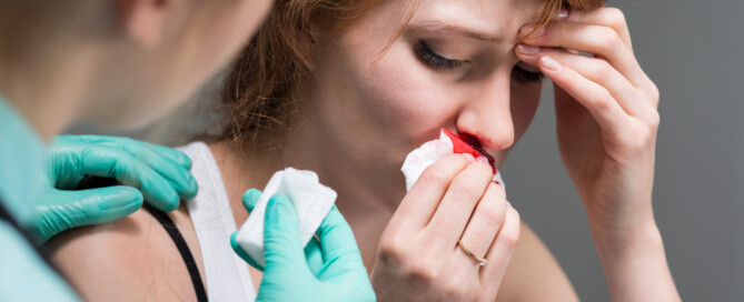
Nosebleeds During Menopause: Hormones or Cancer?

There are many causes of benign nosebleeds, but when they really start up during menopause, a woman may wonder if this means cancer or just the hormonal fluctuations during this transition. (more…)

What Kind of Pituitary Tumor Causes Nipple Discharge?

A certain type of tumor in the pituitary gland can cause nipple discharge by increasing the secretion of prolactin, a hormone. (more…)
What Is Mild Sleep Apnea? It’s Not What You Think

Is the mildness of sleep apnea determined by the patient’s daytime symptoms such as severity? Is it frequency of these symptoms? Or is it something else?

When Is It Okay to Smoke After a Root Canal?

At some point after a root canal, a smoker is going to want to smoke. And at some point following a root canal, it will be okay to smoke.
“Let’s be honest – you’d be hard pressed to find a dentist who thinks ‘smoking is okay’ at any time!” says Gigi Meinecke, DMD, FAGD, member of the Academy of General Dentistry with a private practice in Potomac, MD.
And if your dentist or hygienist is a smoker, this doesn’t change the fact that tobacco is bad for the teeth, gums and everyplace else inside your mouth.
The facts are what they are. But back to the specific question at hand here…
Dr. Meinecke continues, “With that said, there is nothing about a root canal procedure that would be considered a contraindication to smoking.”
So feel free to light up at any point following your root canal.
And maybe right after the procedure you’ll be too stressed to think about kicking the expensive habit.
But at some point, please do yourself a favor and make an effort to dump the habit.
Your teeth, skin and hair will be eternally grateful you did!
And so will your heart and lungs!
 Dr. Meinecke,