How to Convince Elderly Parent to Get Sleep Apnea Test
Here’s how to convince your elderly parent, whom you suspect has obstructive sleep apnea, to agree to a sleep study or an overnight oxygen level test.
I was finally able to convince my advanced-elderly father to e-mail his doctor with a request for a home sleep study. (more…)
Diagnosed with Sleep Apnea in Old Age: Is CPAP Worth It?
Are you elderly and have just been told you have obstructive sleep apnea? Can you still benefit from CPAP therapy? Or should you just use supplemental oxygen?
If your primary care doctor believes you’re too old to benefit from a CPAP machine, and that you should have only supplemental oxygen during sleep, you’ll want to really rethink this recommendation — even if you’re over 90.
“Supplemental oxygen is not a therapy for OSA,” says Dr. Nancy Foldvary-Schaefer, DO, MS, and Director, Sleep Disorders Center, Cleveland Clinic.
“It does not keep the airway open.”
Supplemental oxygen, delivered through a nasal cannula, will elevate the level of oxygen in the blood, and therefore, throughout the entire body.
This may sound like a fix for sleep apnea’s desaturation (abnormally low blood oxygen level).
But it’s far from it. As Dr. Foldvary-Schaefer points out, the oxygen flowing through the nasal cannula does not keep the upper airway open.
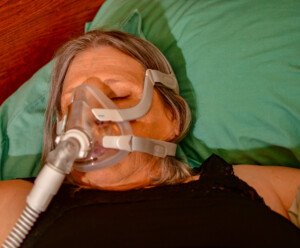
Only the pressurized air from a CPAP hose will do this.
Shutterstock/Grossinger
The airway that keeps getting blocked from collapsed throat tissue — even if supplemental oxygen is keeping the level at 99% — STILL continues to create other problems, including strain on the heart.
“We published a paper in 2019 in JCSM showing that older adults had comparable functional improvement when OSA was treated with CPAP as younger adults,” says Dr. Foldvary-Schaefer.
Another Study Showed the Same Results
A study in a 2015 European Respiratory Journal focused on CPAP benefits for the elderly.
Untreated sleep apnea in a senior man or woman is a whole separate issue from their advanced age, as far as risk or cause for health problems.
There is a distinction, however, in that in the elderly, three complications of untreated sleep apnea are far more likely than in younger patients:
• Frailty
• Nocturia: overnight trips to the bathroom
• Impaired cognition
The conclusion of the European Respiratory Journal paper is that clinicians should not be any less, or more, aggressive in treating obstructive sleep apnea in old people than in middle aged or young patients.
What about long-term benefits of CPAP in an elderly person?
“Benefits in terms of long-term reduction in cardiovascular events is debatable in old adults,” says Dr. Foldvary-Schaefer.
But there are short-term or immediate benefits to consider.
One of them is nocturia. If overnight trips to the bathroom are reduced, as a result of CPAP therapy, this means a reduced risk of falling and breaking a hip or suffering another injury — especially in an elderly person who’s unstable on their feet even when wide awake.
Another short-term benefit is increased alertness during the day, due to restorative sleep.
This means more alert driving and less daytime napping.
Elderly Person’s Oxygen Level During Sleep Is Low; Apneas Are Witnessed by Family Members
My elderly father’s breathing kept pausing for up to 35 seconds during daytime naps. He also snored and frequently fell asleep while watching TV.
An overnight pulse oximeter revealed desaturation, and he was prescribed supplemental oxygen.
I asked his doctor why he wasn’t ordered a sleep study for obstructive sleep apnea, with the idea that CPAP therapy would then be pursued.
His primary care physician replied that Kaiser Permanente didn’t recommend CPAP for suspected sleep apnea cases over 80 because — as Kaiser assumed — people that old would predictably find the face mask to be intolerable — due to physical discomfort on thin old skin, rather than age-related cognitive decline.
“Compliance is only a problem in cognitively impaired patients who do not cooperate,” notes Dr. Foldvary-Schaefer.
Another Study: CPAP Adherence in the Elderly
A paper in the American Journal of Respiratory and Critical Care Medicine (2016) points out (as does the report above) that studies on CPAP use and benefits in the elderly are few.
Neill et al set out to compare CPAP compliance in people 65+ to that of younger patients.
The researchers found that the “mean compliance in the older adults was similar to younger adults.”
However, the paper then states, “Compliance was highest in the > 75 year old group.”
Presence of diabetes or high blood pressure did not affect adherence, either. Nor did home support.
The study concluded that there was a very favorable one-month CPAP compliance in the elderly when it was “delivered as part of a patient focused protocol focusing on the first four weeks of therapy.”
If an elderly family member is suspected of having sleep apnea, encourage them to undergo a sleep study, even if they’re 95.
If they refuse, encourage a pulse oximeter test overnight. If the result shows low oxygen levels, don’t be afraid to discuss a sleep study and the benefits of CPAP therapy.
 Certified by the American Board of Neurology and Psychiatry in Neurology, Clinical Neurophysiology and Sleep Medicine, Dr. Foldvary-Schaefer has treated patients with sleep disorders and epilepsy at Cleveland Clinic since 1995. She has served as a lead investigator on numerous clinical trials.
 Lorra Garrick has been covering medical, fitness and cybersecurity topics for many years, having written thousands of articles for print magazines and websites, including as a ghostwriter. She’s also a former ACE-certified personal trainer.
.
Top image: Depositphotos.com
Sources
erj.ersjournals.com/content/46/1/13
atsjournals.org/doi/abs/10.1164/ajrccm-conference.2016.193.1_MeetingAbstracts.A2851
3.imperial.ac.uk/newsandeventspggrp/imperialcollege/newssummary/news_26-8-2014-17-18-11

Is The Chest Pain of Prinzmetal Angina Always Severe?
Prinzmetal angina chest pain not only usually occurs at rest, but it can be severe.
The severity of chest pain from Prinzmetal angina can be enough to awaken one from sleep. (more…)
Repeatedly Strangled While Sleeping: Sleep Apnea’s Dangers
Don’t think for a second that the typical patient with sleep apnea is a fat man with a thick neck.
Thin young women can be diagnosed with this often misdiagnosed and very common condition.
In fact, it’s estimated that 25 percent of the population has obstructive sleep apnea, and that millions of people go through life having no idea that they have this condition.
Even kids can have obstructive sleep apnea.
Causes are plenty and not always tied to body weight or age.
Causes of this common and under-diagnosed condition include drinking alcohol before bedtime, smoking, large tonsils, large adenoids, a big tongue base, naturally excess throat tissue, a small jaw and a narrow airway.
And of course, obesity and a short thick neck are major risk factors as well.
Obstructive sleep apnea is strongly linked to serious illnesses including heart disease, high blood pressure and stroke.
In addition, “People with sleep apnea are more likely to develop insulin resistance even if not overweight,” says Daniel Rifkin, MD, a sleep medicine expert at the Sleep Medicine Centers of Western New York.
“With this condition, cells don’t respond as well to the hormone insulin, which can lead to type 2 diabetes.
“Sleep apnea can cause decreased oxygen to the optic nerve, which can result in nerve damage.” This can then lead to glaucoma.
“Sleep apnea has been linked to fatty liver disease,” continues Dr. Rifkin. “The severity of the liver disease often corresponds with the severity of the sleep apnea.
“A lot goes on in the brain during sleep. One of those things is the consolidation of memories. With apnea-interrupted sleep, you may have memory problems.”
Untreated sleep apnea mimics other conditions including ADHD, age related cognitive decline, tension headaches and stress.
 Dr. Rifkin is board certified in both neurology and sleep medicine. He also treats insomnia, RLS and narcolepsy.
Lorra Garrick has been covering medical, fitness and cybersecurity topics for many years, having written thousands of articles for print magazines and websites, including as a ghostwriter. She’s also a former ACE-certified personal trainer.
Top image: ©Lorra Garrick
Does Anxiety Really Cause Nocturia or Is Something Else Happening?
If anxiety truly causes nocturia (waking to urinate several times overnight), certainly there has to be a logical reason for this.
You may have read somewhere that stress can increase the urge to void. (more…)
Can You Have Sleep Apnea Without Daytime Symptoms?
Have you been convincing yourself that you can’t possibly have sleep apnea because you don’t have daytime symptoms?
There is a possibility that you have sleep apnea even if you’re not sleepy during the day or experience other daytime symptoms such as morning headaches and trouble concentrating.
When a sleep study (polysomnography) indicates anywhere from five to 15 disrupted breathing events per hour, the diagnosis is mild sleep apnea.
Even in the absence of daytime symptoms.
“Yes, it’s possible,” says Daniel Rifkin, MD, a sleep medicine expert at the Sleep Medicine Centers of Western New York.
“Years ago, we always thought sleep apnea had to be associated with daytime sleepiness.
“In fact, according to certain health insurances, we couldn’t even treat patients unless they were sleepy.
“However, as we learned more and more, we realized that obstructive sleep apnea can have multiple ‘phenotypes’ or presentations.
“Some patients might have mild disease and have profound daytime sleepiness, and others with severe disease and almost no sleepiness at all.
“Those without daytime sleepiness, for example, might have greater cardiovascular risk.”
Compliance with a CPAP Machine
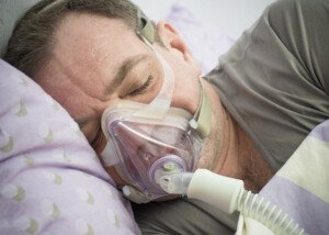
Shutterstock/Oleg Golovnev
If someone has significant daytime symptoms (fatigue, frequent grogginess, often feeling a need to nap, falling asleep on the job), they are more likely to adhere to CPAP therapy, when compared to someone with no daytime symptoms.
Nevertheless, many physicians recommend CPAP therapy for those diagnosed with even mild obstructive sleep apnea even in the absence of daytime symptoms.
Other sleep medicine doctors question the value of CPAP for those with asymptomatic mild obstructive sleep apnea — if for no other reason, there’s less likely to be compliance in such individuals.
Adherence should be taken into account, says a paper in the Journal of Clinical Medicine.
AHI is the apnea hypopnea index. Its associated numerical value refers to the average number of these events per hour of sleep.
Hypopnea refers to inhaling against resistance created by a partially rather than completely obstructed airway.
An AHI of five to 15 means mild sleep apnea.
Over 15 to 30 is moderate OSA, and over 30 is severe OSA.
Someone with mild OSA is not likely to be compliant with CPAP.
It is not known whether the cardiovascular risk of untreated mild OSA — for which there are no daytime symptoms — is lower than for untreated mild OSA with daytime symptoms.
People with asymptomatic mild OSA may abandon the idea of CPAP therapy once they learn that studies about cardiovascular risk in untreated mild OSA are not conclusive.
The JCM paper states that cohort studies of death rates with CPAP therapy suggest that only those individuals with an AHI greater than 30 (severe OSA) have a reduced mortality.
The JCM also points out that for mild OSA with daytime symptoms, non-CPAP alternatives may be effective.
These include weight loss, changing sleep position and a mandibular advancement device custom made by a dentist.
Dr. Rifkin is board certified in both neurology and sleep medicine. He also treats insomnia, RLS and narcolepsy.
Lorra Garrick has been covering medical, fitness and cybersecurity topics for many years, having written thousands of articles for print magazines and websites, including as a ghostwriter. She’s also a former ACE-certified personal trainer.
.
Top image: Depositphotos.com
Source: ncbi.nlm.nih.gov/pmc/articles/PMC2564770/
Can Untreated Mild Sleep Apnea Cause Cardiovascular Harm?

Don’t let the diagnosis of “mild” fool you; untreated though mild obstructive sleep apnea can lead to adverse cardiovascular events in your body. (more…)
Is Peeling Skin on the Feet a Sign of Diabetes?
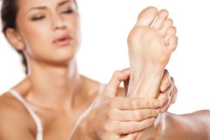
All sorts of things can go wrong with the feet from diabetes and this includes peeling of the skin.
You probably already know that diabetes often causes neuropathy (nerve damage), which results in the inability to feel pain in the foot. (more…)
How to Tell You Might Have Obstructive Sleep Apnea ??


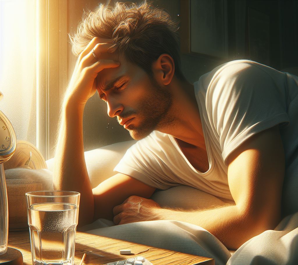
Don’t count yourself out from having sleep apnea just because you don’t have daytime symptoms and are lean, buff and young.
Sleep apnea is one of those conditions for which millions of people could be walking around totally clueless that they have it. Including women.
Classic Symptoms of Sleep Apnea
“Snoring (especially loud enough to disturb others), unrestful sleep, morning headaches, daytime sleepiness (especially if you fall asleep inappropriately such as while driving, talking with others, at work) and of course, fatigue,” says Susan L. Besser, MD, with Mercy Medical Center, Baltimore; Diplomate, American Board of Obesity Medicine and board certified by the American Board of Family Medicine.
The person with untreated sleep apnea may be prone to napping a lot but never feeling recharged.
He or she may avoid extended driving trips because they know they’ll become drowsy behind the wheel.
The morning headaches disappear soon after getting out of bed.
You may not have the more overt signs of sleep apnea, such as snoring or excess daytime grogginess.
However, there are nighttime signs that are suspect for untreated sleep apnea.
• Suddenly awakening feeling as though your airway was cut off
• Partner describes snoring as a snorting sound
• Partner describes your breathing as labored or gasping
• Partner says you stop breathing for extended periods
• Partner says your breathing is erratic, shallow and/or comes in rapid waves between long pauses
• Repeated trips to the toilet overnight
If any of the above bullet points applies to you, you should discuss these with your physician and consider the possibility of having a sleep study — which can determine if you have sleep apnea.
In addition to the above points, do any of the following apply to you?
• Scalloped tongue*
• Can’t see throat due to high-arched palate
• Crowded teeth
• Wisdom teeth removal
• Small appearing jaw
• Receding chin
*Scalloped tongue. Check the sides of your tongue. Do you see indentations from your teeth? That indicates a small jaw.
Your dentist can discuss with you the likelihood of sleep apnea if you have any of the oral features listed above.
Dentists view the interior of mouths all day long and can give you very insightful feedback.
 Dr. Besser provides comprehensive family care, treating common and acute primary conditions like diabetes and hypertension. Her ongoing approach allows her the opportunity to provide accurate and critical diagnoses of more complex conditions and disorders.
 Lorra Garrick is a former personal trainer certified through the American Council on Exercise. At Bally Total Fitness she trained women and men of all ages for fat loss, muscle building, fitness and improved health.
What Is the Black Debris in Gum Where Tooth Was Removed?