You know what an MRA is, but what exactly happens during an MRA – here is my firsthand account.
I had an MRA – no, not MRI, but MRA, as in magnetic resonance angiogram.
An MRA is a type of MRI, actually.
Perhaps your doctor has told you that you should have an MRA.
Or maybe you’ve already made the appointment for the magnetic resonance angiogram.
There are many reasons to have an MRA, and just because your doctor has ordered this procedure, doesn’t mean it will find something wrong with you.
What’s an MRA like?
After filling in the paperwork, you’ll wait.
The technician who did my procedure was the person who came into the waiting room to summon me.
I was led to a room and told to slip into two gowns (I don’t remember the reason why two were necessary, but it made sense at the time).
I then waited in an area for people waiting to get an IV portal inserted into their arm for the contrast dye.
What happens during an MRA?
The procedure uses contrast dye to make the blood vessels visible. So I waited briefly, then was called into a room where a nurse prepped me for the IV portal.
Then I was sent back to the little waiting room.
The technician then summoned me. I had asked for a chance to speak to the cardiologist who’d be present.
I was also told I couldn’t wear earplugs (magnetic resonance procedures produce loud noise) because I had to wear headphones to be able to communicate with the technician during the exam.
I briefly spoke to the cardiologist, and then was taken to the MRA room.
I lied on the table. The technician placed some heart monitor leads on my chest.
I had been told that during the procedure I’d be having to hold my breath, usually for 20 seconds at a time.
The technician placed a thick, weighty rectangular-shaped thing on top of my chest, and did something with the IV portal, though she did not inject the dye.
The dye is injected from another room. I don’t know how this works, but after I had lied on the table, she had rigged something with the IV portal, while I was lying flat with my eyes closed.
I was told the test would take an hour, more likely an hour and 15 minutes.
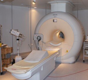
A little hand pump was placed in my other hand and I was told that if I needed to be taken out of the MR tube, to squeeze the pump.
The MRA was of my heart (I’ll get to the reason in a moment), so my head was completely in the tube.
Many times I was told (through the headphones by the tech) to “Inhale, exhale, now take a deep breath and hold.” Then she’d say, “Breathe.”
This pretty much defined the experience, though there were several points during which she told me to relax for a few minutes while she made adjustments from the other room.
Believe it or not, only the last few minutes of the procedure involved the contrast dye.
She alerted me when she was giving it to me (while I was still inside the tube). I had no reaction to it. Next thing I knew, the exam was done.
Why did I have the MRA? I had had a visit with a cardiologist as part of heart disease screening (both parents have heart disease).
He detected a Class II heart murmur. The subsequent echocardiogram was normal, so the only thing he could think of that was causing the murmur was pulmonary stenosis (narrowing of pulmonary artery). A magnetic resonance angiogram would detect this.
In my case, the MRA turned out completely normal, and my cardiologist has concluded that the murmur is a “functional flow” type which can develop in athletes (of which I am). It is harmless.
I had my next routine screening two years later, and the cardiologist said, “You have the smallest heart murmur in the world. I don’t hear it.”

