

Can Sinus Headache Come from Trigeminal Nerve Irritation?

All kinds of things can cause a headache, but what about the trigeminal nerve which is in the nasal passages (sinuses)?
Can this nerve cause a referral type of pain into the head?
“Trigeminal nerve irritation can cause headache,” says Dr. Stacey Silvers, MD, of Madison ENT & Facial Plastic Surgery in NYC, who is board certified in otolaryngology; one of her specialties is sinus surgery.
Dr. Silvers continues, “Patients with trigeminal neuralgia have classic pain anywhere along the course of the trigeminal nerve. There are branches that go as high as the temple, causing sharp stabbing pains on the sides of the head.”
The discomfort can also feel more like a tension type of headache — the kind that’s caused by a stressful situation.
“Though trigeminal nerve headaches are different from migraine and tension varieties, chronic pain can cause chronic tension and stress which can lead to headache,” says Dr. Silvers.
“Migraines can have a variety of triggers (loud noises, flashing lights, foods and even chronic pain).
“People can have headaches from the trigeminal nerve itself based on its course through the skull, or the resulting tension or migraine headache that the neuralgia (chronic pain) may trigger,” due to the emotional stress of having this condition.
Aggressive cleaning of the nose by putting objects up the nasal passages such as a cotton swab can irritate the trigeminal nerve, which has branches extending into the nasal passageways low enough for a cotton swab or finger to easily reach.
Over time, repeated scooping out of mucus chunks can start irritating the trigeminal nerve, leading to headaches.
The solution is to irrigate the nose with a neti pot; this will soak the hardened mucus so that it can be blown out, or, if it’s still stubborn, at least it can be more gently scooped out.
 An NYC expert in ear, nose and throat care, Dr. Silvers has been named among America’s Top Physicians and Surgeons in facial plastic surgery and otolaryngology numerous times since 2003. Dr. Silvers is an expert in the field of minimally invasive rhinology, resolving patients’ breathing and sinus problems with simple in-office procedures.
 Lorra Garrick has been covering medical, fitness and cybersecurity topics for many years, having written thousands of articles for print magazines and websites, including as a ghostwriter. She’s also a former ACE-certified personal trainer.
.
Top image: ©Lorra Garrick

Can a Nasal Polyp Irritate the Trigeminal Nerve?

Is it possible for a nasal polyp to irritate the trigeminal nerve, being that this nerve extends into the nasal passage?
“Trigeminal neuralgia is chronic irritation or pressure on the trigeminal nerve,” says Dr. Stacey Silvers, MD, of Madison ENT & Facial Plastic Surgery in NYC, who is board certified in otolaryngology; one of her specialties is sinus surgery.
“It can be caused by compression of the nerve by a mass, trauma directly to the nerve or effects on the blood flow supplying the trigeminal nerve,” continues Dr. Silvers.
“Polyps do not cause pressure and they themselves have no sensation; the mucosa that they are attached to is where the sensation is. Polyps are not known to irritate the trigeminal nerve.”
A nasal polyp is a soft, painless and benign growth that usually occurs in adults, though they can develop in people of all ages. They cannot turn into cancer.
Often, these growths are discovered during a routine exam when a doctor peers up the patient’s nose. Small growths typically do not cause symptoms.
Nasal polyps can cause quite a few symptoms.
-Persistent stuffiness in the absence of a cold
-Runny nose despite no cold or allergy
-Postnasal drip despite no cold or allergy
-Loss of sense of taste
-Diminished or total loss of smell
-Headache or facial pain
-A pressure feeling over the forehead or face
-Pain in the upper teeth
-Itching around the eyes
-Snoring
If you have any of these symptoms and a doctor has not determined causation, then have an otolaryngologist examine you for possible nasal polyps.
Treatment is either with surgery or medication. These benign growths can grow back after being successfully treated.
Regarding the trigeminal, Dr. Silvers adds, “The nerve does have branches in the nose, and rare reports of trigeminal neuralgia have been seen after aggressive and minimally invasive sinus/nasal surgery.”
An NYC expert in ear, nose and throat care, Dr. Silvers has been named among America’s Top Physicians and Surgeons in facial plastic surgery and otolaryngology numerous times since 2003. Dr. Silvers is an expert in the field of minimally invasive rhinology, resolving patients’ breathing and sinus problems with simple in-office procedures.
Lorra Garrick has been covering medical, fitness and cybersecurity topics for many years, having written thousands of articles for print magazines and websites, including as a ghostwriter. She’s also a former ACE-certified personal trainer.

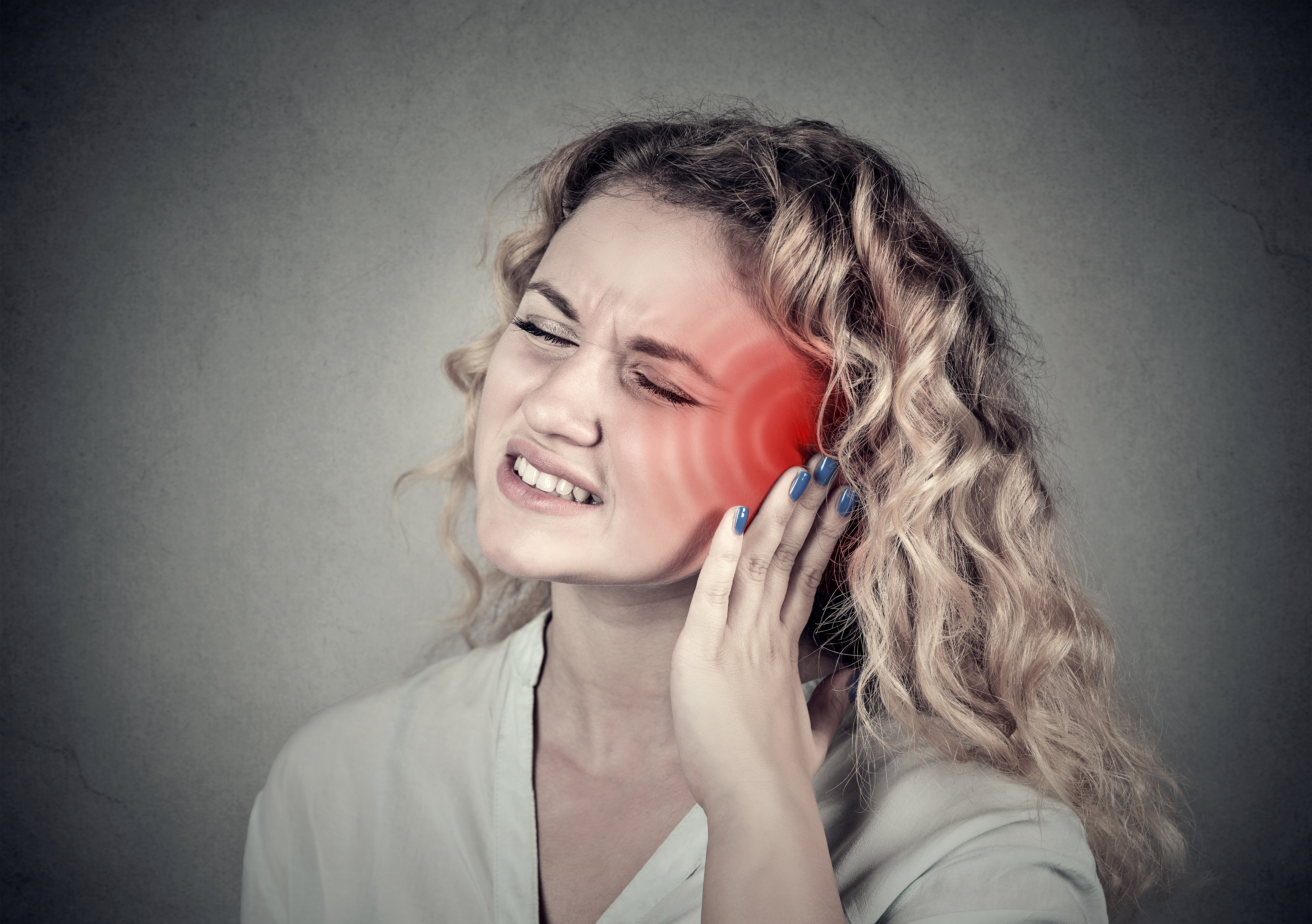
Can Nasal Polyps Cause Ear Pain?

Ear pain can be caused by a nasal polyp.
“Nasal polyps can cause nasal congestion and rhinitis with nasal mucous,” says Dr. Stacey Silvers, MD, of Madison ENT & Facial Plastic Surgery in NYC, who is board certified in otolaryngology; one of her specialties is sinus surgery.
“They develop usually from an allergic reaction to an environmental irritant,” continues Dr. Silvers.
“They are found commonly when we look with a scope in the nose, and in many patients are not symptomatic and found on routine exam.
“Some patients are very symptomatic and the polyps will affect breathing, sense of smell and block the sinuses.”
If you seem to be having difficulty breathing through your nose, it may very well be a polyp, which can be surgically removed, or, more conservatively treated with medication.
These potentially bothersome growths are non-cancerous, and they cannot turn into cancer. They can return even after disappearing from successful treatment.
“Nasal congestion can commonly cause ear congestion or blockage of the Eustachian tube.”
Dr. Silvers adds that ET congestion causes clogging as well as pressure in the ear, and “sometimes pain in the ear.
“Polyps are not typically seen blocking the Eustachian tube, but the congestion that is associated with polyps that affects the surrounding mucosa can cause these ear symptoms.”
So yes, a nasal polyp can definitely cause ear pain. However, ear pain can have many causes, including having recent aggressive cleaning with a cotton swab as well as a condition called Eagle’s syndrome.
An NYC expert in ear, nose and throat care, Dr. Silvers has been named among America’s Top Physicians and Surgeons in facial plastic surgery and otolaryngology numerous times since 2003. Dr. Silvers is an expert in the field of minimally invasive rhinology, resolving patients’ breathing and sinus problems with simple in-office procedures.
Lorra Garrick has been covering medical, fitness and cybersecurity topics for many years, having written thousands of articles for print magazines and websites, including as a ghostwriter. She’s also a former ACE-certified personal trainer.
.
Top image: Shutterstock/pathdoc

Why Coughing Every Several Minutes Can Be Caused by Anxiety

You may already know that anxiety can cause frequent coughing, but do you know just how this happens?
Perhaps your coughing is similar to what I experienced during a horrible time of my life.
At some point amid tremendous anxiety, I developed a cough.
I had to cough literally every several minutes due to an itchy, tickly feeing in the back of my throat that I couldn’t ignore.
I’d try to resist coughing, but sooner or later, the urge to clear up that itching and tickling became too strong.
Often, the cough would bring up what appeared to be tiny droplets of saliva.
This went on for several days and I knew it was somehow related to my anxiety. I had no other symptoms that would normally go with a cough.
During the times I was transfixed on my computer with deadline work, I noticed that the cough was suppressed. I also never had it while in bed or during exercise.
One of my stressors was a pending colonoscopy because I was experiencing sudden onset diarrhea and a change in stool caliber that was leaving me dehydrated and thinking colon cancer.
Two days before the colonoscopy, my beloved dog had to be euthanized after we incorrectly thought his brain cancer was retreating.
He had quickly taken a turn for the worse and became deranged three days before the colonoscopy. The stress of everything was unbearable.
The coughing every several minutes accompanied me straight to the colonoscopy procedure room until I lied down.
When the procedure was over, I sat, the doctor told me everything looked normal, the sedatives rapidly wore off, and I realized that the cough was gone. And I mean completely gone. It never returned.
Stacey Silvers, MD
“Your stress, like many others, can cause acid reflux,” begins Dr. Stacey Silvers, MD, of Madison ENT & Facial Plastic Surgery in NYC, who is board certified in otolaryngology (ear, nose and throat).
“Stress increases stomach acid; this acid refluxes up into the back of the throat.
“The result is: postnasal drip to coat the throat from the acid, and a swelling around the opening of the esophogus.
“That swelling will tickle the vocal cords and cause a chronic dry cough and throat clearing.
“Ninety percent of all patients presenting with these symptoms have a stressful event or more than one that occurred when the symptoms started.
“Postnasal drip is only a reaction to the reflux and acts as a protector of the throat from the stomach acid which is a pH of 2.
“This mucous and postnasal drip is why people do not have classic burning symptoms.
“The postnasal drip and resulting swelling from the reflux can trigger a cough, however.
“It is not from the sinuses or from sudden onset of allergies. It is very common and very commonly misdiagnosed and mistreated.
“When the stressor goes away (normal medical study, successful move, successful job change or adjustment to the death of a loved one or pet), the symptoms often go away.
“During the symptoms, however, reflux medication will stop the postnasal drip and therefore cough.”
If you find that you’re coughing every several minutes but don’t have accompanying cold or allergy symptoms, ask yourself if you’ve been under an acute attack of severe anxiety.
An NYC expert in ear, nose and throat care, Dr. Silvers has been named among America’s Top Physicians and Surgeons in facial plastic surgery and otolaryngology numerous times since 2003. Dr. Silvers is an expert in the field of minimally invasive rhinology, resolving patients’ breathing and sinus problems with simple in-office procedures.
Lorra Garrick has been covering medical, fitness and cybersecurity topics for many years, having written thousands of articles for print magazines and websites, including as a ghostwriter. She’s also a former ACE-certified personal trainer.
.
Top image: Shutterstock/Aaron Amat

Can Constant Stress & Anxiety Make You Lose Weight?

You’ve heard that stress can interfere with weight loss efforts, but can a lot of anxiety cause fat loss even if you don’t eat less?
It’s a no-brainer that weight loss will occur from eating less due to suppressed appetite from stress.
“Nobody lives life completely devoid of stress; however, there are times in all of our lives when stress and the associated anxiety that can accompany a significantly greater stressful situation than usual, can be more taxing than we even realize,” says Richard Kelley, MD, a practicing physician in Texas for 20+ years, and author of “The Fitness Response,” “The Three-Hour Appetite” and the ebook, “The Fitness Response ‘Diet’ for Women.”
He continues, “Prolonged stress and anxiety can take a significant toll on one’s health, inducing weight loss, insomnia, gastrointestinal problems and general irritability.”
“Weight loss as a result of increased stress and anxiety may occur for a variety of reasons.
“When we are chronically stressed and anxious, whether we realize it or not, nutrition may suffer due to skipping meals.
“For individuals caring for an ill family member for extended periods of time, either at home or in the hospital, one’s sense of time can be altered, resulting in skipped meals or prolonged periods between nutrition.
“It is not unusual for a family member in the chronic caretaker role, to feel they are eating adequately and even have an increased sense of hunger when they do eat, simply because their schedule, secondary to their caretaking role, is more erratic.”
I had unexplained weight loss while caring for my mother following her quintuple bypass surgery.
However, I was acutely aware of my eating habits simply because I’m a health-conscious person, always striving to increase my muscle mass.

When I began noticing the weight loss, I began tracking caloric intake, even though I knew I was eating more than usual (e.g., many thick personal pizzas at the hospital; my mother had four more hospital admissions after the initial one).
There were three or four days at the beginning where I didn’t eat much, but the rest of the days I ate generously, including a rich homemade chocolate milkshake every day, but lost fat rather quickly.
“Individuals who maintain a fitness-oriented lifestyle and regularly stimulate and activate their skeletal muscle, may find that they lose weight (body fat) over months in a stressful situation, eating even more calories than usual, simply because their focus has been on maintaining their skeletal muscle through their regular routine of resistance training, prior to the period of prolonged stress,” says Dr. Kelley.
“Many people disregard the power and impact of a well maintained musculoskeletal system, but muscle plays a significant role in helping people burn calories, often in the presence of increased caloric intake.”
But why the sudden fat loss in my case?
After all, my body already had the muscle development.
“Prolonged anxiety, in and of itself, may result in a tendency to lose weight,” says Dr. Kelley.
“Anxiety may enhance adrenal activity, producing higher levels of hormones such as adrenaline and cortisol, which increase heart rate and blood pressure.
“This would also represent an elevation in metabolic rate, which over time, could result in the loss of weight.
“Elevated cortisol levels have traditionally been viewed as increasing one’s tendency to store abdominal fat.
“Its role, if any, in the weight loss process, as a result of prolonged stress, is not well-understood.
“Though the argument could be made that prolonged stress and anxiety result in a more consistent elevation of adrenaline than cortisol, over time, or that the impact of the fight or flight hormones simply exert more visible impact (weight loss) over the course of time, than that of cortisol.”
Dr. Kelley also points out (though this didn’t apply to my case) that stressed caregivers sometimes subsist largely on coffee, soda and cigarettes to stay alert, and these substances elevate adrenaline and suppress appetite.
“Ultimately, I believe prolonged stress and anxiety may have a tendency to skew our view concerning the accuracy of our nutrition recall.
“In my own weight management practice, I can say with certainty that most individuals have very poor recall of exactly what and how they have eaten, over the course of several days, a week or a month, if they don’t journal their food intake.
“We are even less likely to fully appreciate how well we are doing, nutritionally, when we are in a state of chronic, prolonged stress.”
In conclusion, a sudden bout of extreme stress can cause weight loss even if you don’t eat less; even if you eat more; and especially if you already have a trained body.
 Richard Kelley, MD, is an author, speaker, fitness expert and transformation coach.
 Lorra Garrick has been covering medical, fitness and cybersecurity topics for many years, having written thousands of articles for print magazines and websites, including as a ghostwriter. She’s also a former ACE-certified personal trainer.

Nighttime Binge Eating, Feeling Hot Overnight & Next Morning

Ever eat a lot of food at night, then get really hot in the middle of the night or next morning?
More than once I’ve had the experience of eating quite a bit of food at night, only to awaken in the middle of the night feeling overheated, or, if not, then awakening the following morning hot — despite a normal room temperature.
These were not hot flashes; just a hot feeling. Intuitively I knew it was because all that extra food made my body work extra hard to digest it — work generates heat.
“Though not a complaint by everyone, the answer to why some individuals experience an increased sensation of heat, after an evening food binge, can be explained, at least to some degree, by the fact that the processing, breakdown and utilization of food, is an energy expending process of the body,” explains Richard Kelley, MD, a practicing physician in Texas for 20+ years, and author of “The Fitness Response,” “The Three-Hour Appetite” and the ebook, “The Fitness Response ‘Diet’ for Women.”
Dr. Kelley continues, “In fact, it is estimated that the processing, breakdown and utilization of ingested food, accounts for approximately 10 percent of the body’s daily energy expenditure.
“This is actually a calorie burning process of the body and can also result in thermogenesis or the generation of heat, by the body.
“The reason that some individuals report more awareness of this phenomenon, while other individuals are less aware of the increase in heat, is not entirely clear.”
Eating a lot causes the body to burn more calories = more heat generated.
If you binge at night, a feeling of heat will be more noticeable because the thermogenesis occurs while you’re lying in bed.
If the generation of heat occurs mid-day, after a morning binge, you’re less likely to be aware of feeling hot, since you’re already active; or, you may feel hot but think nothing of it if you’re concurrently moving about busily.
Dr. Kelley continues, “For an individual who engages in a true binge or excessive caloric intake at one sitting, the body is literally going into overdrive, metabolically speaking, to break down and store the excess of calories.
“That some individuals are sensitive to the fact that metabolism, along with thermogenesis, is increasing as a result of the binge, is not surprising, given that we know the body literally has to get to work anytime we put food into the stomach, and this is certainly the case when we overfeed ourselves.”
Next time you binge at night and then several hours later feel hot, you now know why.
Richard Kelley, MD, is an author, speaker, fitness expert and transformation coach.
Lorra Garrick has been covering medical, fitness and cybersecurity topics for many years, having written thousands of articles for print magazines and websites, including as a ghostwriter. She’s also a former ACE-certified personal trainer.

Causes of Armpit Lumps other than Cancer

A lump in you armpit doesn’t necessarily mean cancer, because it more likely has a benign cause.
Have you recently discovered a lump in your armpit and immediately thought, “It’s cancer!”?
Relax, because chances are in your favor that the lump under your arm does not have a life-threatening cause.
It’s just that the discovery of a lump — whether visible or only detectable with probing fingers — is one of the warning signs that we’ve all learned can be caused by many different cancers.
Cancers that are infamous for causing lumps that are first detected by the patient include that of the breast, prostate, testicles, lymph nodes, bone and inside the mouth.
Origin of Most Underarm Lumps
“In general, armpit lumps are from lymph glands or sweat glands,” says Dr. Steven Lamm, MD of internal medicine, who appears regularly as the house doctor on ABC’s “The View,” and author of “No Guts, No Glory,” a book about digestive issues.
Dr. Lamm continues, “Lymph glands can swell from infections anywhere in the arm or from a generalized infection.
“Sweat glands, also, can get blocked and swollen. The most common condition is called hidradenitis.”
Hidradenitis suppurativa is a chronic skin inflammatory condition that can last for years, characterized by blackheads and at least one tender bump that can get bigger, break open and ooze pus.
The armpits and groin are common locations. Finally, a cause of a “lump” under the arm can simply be your natural lymph node.
Some years ago while performing a self-breast exam I found a lump under my arm, and could not locate a mirror image of it on the other side—initially, that is.
Eventually I was able to feel around my other armpit and locate a similar lump in the same relative location.
Variations in sinew and muscle can account for why, on one side, there seems to be a “lump,” while on the other side, there’s none there.
 On the clinical faculty in internal medicine at New York University Medical Center, Dr. Lamm has maintained a private practice in NYC for 30+ years.
Lorra Garrick has been covering medical, fitness and cybersecurity topics for many years, having written thousands of articles for print magazines and websites, including as a ghostwriter. She’s also a former ACE-certified personal trainer.
.
Top image: Freepik/valuavitaly

Why Vomiting Can Be Triggered by Anxiety

Are you thinking that anxiety causes vomiting because it’s the body’s way of unloading something useless for the imminent “flight or fight” situation?
Anxiety can be chronic or acute. People will vomit upon seeing a mashed raccoon on the road (acute stress) or periodically upchuck from the stress caused by battling an ex for custody of the kids.
So it’s no secret that some people will vomit during intense episodes of anxiety, but what’s the mechanism behind this?
Have you ever known anyone who vomited as a result of a lot of anxiety?
Of course, people have been known to upchuck even upon smelling something horrible such as rotten fish.
Anxiety, Stress and the Gut

Freepik.com, Diana.grytsku
“Anxiety causes the brain to send a signal to the stomach through the vagus nerve, causing changes in the physiology of the gut,” says Dr. Steven Lamm, MD, of internal medicine, who appears regularly as the house doctor on ABC’s “The View,” and author of “No Guts, No Glory,” a book about digestive issues..
Dr. Lamm continues, “In addition, anxiety causes changes in the center of the brain involved with nausea as well.
“It’s not uncommon for an anxious person to feel they need to vomit. But this is with extreme anxiety.”
An exercise regimen can help combat the physical effects of chronic anxiety.
Ideally, an exercise program should consist of both aerobic activity and weight bearing activity.
Yoga is another form of exercise that can help a person manage anxiety and its fallout including nausea and vomiting.
On the clinical faculty in internal medicine at New York University Medical Center, Dr. Lamm has maintained a private practice in NYC for 30+ years.
Lorra Garrick has been covering medical, fitness and cybersecurity topics for many years, having written thousands of articles for print magazines and websites, including as a ghostwriter. She’s also a former ACE-certified personal trainer.
.
Top image: ©Lorra Garrick

Can Mononucleosis Cause Bleeding in the Throat?

Mononucleosis indeed can cause a bloody throat, says a physician.
What should you think if you look into your teenager’s throat and see blood?
Mononucleosis wouldn’t be the first thing that came to your mind if your child, or you yourself, had bleeding in the throat, unless you already suspected this viral infection.
My niece has mononucleosis and at one point, her mother peered into her throat and saw what was described as “bloody.”
So I asked a physician if this can be a consequence of this illness.
“Yes, mono is a viral fungus causing significant inflammation of the tonsils,” says Dr. Steven Lamm, MD, of internal medicine, who appears regularly as the house doctor on ABC’s “The View,” and author of “No Guts, No Glory,” a book about digestive issues.
Dr. Lamm adds, “Severe inflammation can actually cause inflammation to the point of bleeding. This, however, is very rare.”
According to mayoclinic.com, here are the more common symptoms of this infection (bleeding is not listed):
Sore throat, swollen tonsils, fatigue and weakness, fever, swollen lymph nodes in the armpits and neck, skin rash, headache, loss of appetite, swollen spleen and night sweats.
Mayoclinic.com says that the incubation for the virus is four to eight weeks, but in kids it can be shorter.
The reason mononucleosis is dubbed “the kissing disease” is because it’s spread through saliva.
Though blood in the throat can be scary, this does not mean a serious complication.
On the clinical faculty in internal medicine at New York University Medical Center, Dr. Lamm has maintained a private practice in NYC for 30+ years.
Lorra Garrick has been covering medical, fitness and cybersecurity topics for many years, having written thousands of articles for print magazines and websites, including as a ghostwriter. She’s also a former ACE-certified personal trainer.
.
Top image: Shutterstock/Prostock-studio

How Long Can Chest Pain from a Panic Attack Last?

The chest pain that comes from a panic attack can last surprisingly long, tricking you into thinking you’re having a heart attack in progress.
On the other hand, a panic attack may cause chest pain that lasts only a very short period of time.
A panic attack is a sudden episode of intense fear or discomfort, often accompanied by physical symptoms like rapid heartbeat, pain in the chest, sweating, trembling and shortness of breath.
It can be overwhelming and may feel like you’re losing control or having a heart attack.
How long can chest pain from a panic attack actually last?
“Minutes to hours,” says Dr. Steven Lamm, MD, of internal medicine, who appears regularly as the house doctor on ABC’s “The View,” and author of “No Guts, No Glory,” a book about digestive issues.
“It can certainly be confused with heart attack,” adds Dr. Lamm.
Chest pain from coronary artery disease (in the absence of an imminent heart attack), as well as from severely blocked coronary arteries in which a heart attack is imminent, can last a few minutes to a few hours.
“From coronary disease, it’s generally initiated by exercise and relieved by rest,” says Dr. Lamm.
In the case of my mother, prior to her quintuple bypass surgery, chest discomfort would be triggered by a few minutes of housework, then relieved upon sitting down.
However, two days before her surgery, the chest pain occurred while she was sleeping. It had awakened her and persisted for two hours.
Something to consider: What if you’re having chest pain or some kind of discomfort in that region, but you do not feel panicky or are not behaving or thinking in a way that’s classic for a panic attack?
For instance, a sudden, intense feeling of being out of control and that death is moments away are a few classic features.
So if you’re minus these hallmark symptoms of a panic attack, you just might be having a problem with your heart.
Dr. Lamm continues, “Panic disorders often occur at rest and can theoretically diminish w/exercise.”
If you feel a panic event coming on, do some pushups or jumping jacks and see what happens. I can speak from experience.
I’ve had more than one panic attack (no trigger), and for one of the events, I did pushups and the episode quickly disappeared.
Dr. Lamm says that the chest pain with coronary artery disease has a crescendo pattern.
This means that the pain steadily gets worse and worse when it occurs. “With panic, it’s generally a steady pain that can come and go.”
On the clinical faculty in internal medicine at New York University Medical Center, Dr. Lamm has maintained a private practice in NYC for 30+ years.
Lorra Garrick has been covering medical, fitness and cybersecurity topics for many years, having written thousands of articles for print magazines and websites, including as a ghostwriter. She’s also a former ACE-certified personal trainer.
.


