

How Soon after a Normal Head CT Can You Drive?

It’s a fair question: For how long should you avoid driving after hitting your head, even though the CT scan is normal and you have no symptoms?
You hit your head and that day, get a CT scan to see if there’s any sign of subdural hematoma. The CT scan is normal.
However, you also know that a normal CT scan the day of head trauma, and even 24 hours later, doesn’t guarantee you can’t develop a slow brain bleed (chronic subdural hematoma) over the next few months…causing disturbing neurological symptoms.
Some of the symptoms that a chronic subdural hematoma can cause are sudden and profound weakness of one or both legs or arms; a seizure; cognitive impairment; and a searing headache.
In fact, a symptom from chronic subdural hematoma can mimic that of stroke or transient ischemic attack. Imagine driving while a symptom (or two) suddenly strikes.
Could you control the car while your brain is bleeding?
You may not even KNOW you’re having a symptom, and thus, begin losing control of the car and not even know it!
This begs the question:
For how long should a person (particularly elderly) avoid driving even if their CT scan (and next-day follow-up CT scan) is normal, and they are sent home with no symptoms?
“It depends on severity of initial presentation,” says Ivan Mikolaenko, MD, a board certified neurologist of 20+ years based in New York with subspecialty certification in neurocritical care.
“I usually recommend to take a rest and don’t drive until all symptoms are gone, and I usually recommend to repeat CT scan in 3-4 weeks after head injury in patients older than 60 years.
“After that, given normal exam and normal imaging, patient can go back to driving.”
In other words, no symptoms at initial presentation in ER, and with a normal CT scan, mean that the patient is free to drive.
However, the patient should never drive TO the ER after hitting their head, since at that point, no diagnosis has been made.
You should always have someone drive you to the ER even if you “feel fine.”

Conditions treated by Dr. Mikolaenko in the intensive care setting include strokes, ruptured aneurysms, trauma-related brain and spinal cord injuries, seizures and brain swelling, infections and tumors.
 Lorra Garrick has been covering medical, fitness and cybersecurity topics for many years, having written thousands of articles for print magazines and websites, including as a ghostwriter. She’s also a former ACE-certified personal trainer.
.
Top image: ©Lorra Garrick

Mortality of Chronic Subdural Hematoma Soon After Drainage?

A certain percentage of people will die soon after having their chronic subdural hematoma drained.
The drainage procedure takes only 15 to 20 minutes, yet within 30 days, five percent of patients will be dead.
A 5% Mortality Rate within 30 Days of Burr Hole Drainage of a Chronic Subdural Hematoma
Just prior to my mother undergoing the 15-20 minute drainage procedure for her chronic subdural hematoma, the neurosurgeon told me that the biggest risks were infection and recurrence.
He made no mention of a mortality rate for the noninvasive procedure.
However, I’ve read in several online neurosurgery journals that there’s a well-documented mortality rate in patients within 30 days of the burr hole drainage of their chronic subdural hematoma.
One study said a 5 percent mortality rate. Another said a 6 percent mortality rate out of the 157 patients investigated.
“Although most of cSDHs often appear innocuous, severe complications may occur,” says Ivan Mikolaenko, MD, a board certified neurologist with subspecialty certification in neurocritical care, Neurological Surgery, P.C., New York.
Dr. Mikolaenko says those include the following.
“- Devastating intracerebral hemorrhage due to rapid brain decompression leading to hyperemia (increased blood flow) immediately beneath the hematoma or at any other area of the brain which was shifted and compressed for many days before surgery.
“- Failure of the brain to re-expand, especially in patients with pre-surgery low subdural fluid collection pressure: These people very often will have re-accumulation of the subdural fluid.
“- Intractable seizure post-op with or without status epilepticus.
“- Longstanding cSDH with conversion to subdural empyema (collection of subdural pus).
“- Untreated severe pneumocephalus (collection of bubble of air in place of evacuated subdural collection).”
As frightening as this mortality information all sounds to anyone who’s been diagnosed with a cSDH, it’s very important to realize that the death rate is still relatively low.
If you’ve hit your head recently, or know someone who has, be on the very vigilant lookout for any new-onset suspicious symptoms.
This includes out-of-character change in mood, memory issues, weakness, change in gait pattern, apathy, sudden headaches, or anything that just doesn’t seem right with the individual.
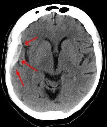
A chronic subdural hematoma is a slow bleeding in the brain, beneath the dura mater, and may take up to 90 days to begin producing symptoms.
Conditions treated by Dr. Mikolaenko in the intensive care setting include strokes, ruptured aneurysms, trauma-related brain and spinal cord injuries, seizures and brain swelling, infections and tumors.
Lorra Garrick has been covering medical, fitness and cybersecurity topics for many years, having written thousands of articles for print magazines and websites, including as a ghostwriter. She’s also a former ACE-certified personal trainer.
.
Top image: Shutterstock/Volodymyr Baleha
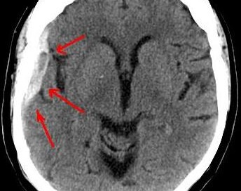
Chronic Subdural Hematoma Questions Answered by Doctor
Here is where you may find the answers to your symptom questions about chronic subdural hematoma—right from a neurosurgeon.
Here are three questions about chronic subdural hematoma symptoms answered by Ivan Mikolaenko, MD, a board certified neurologist of 20+ years based in New York with subspecialty certification in neurocritical care.
How much time must pass (“incubation period” or “grace period”) after the head trauma before the patient can know he won’t develop a chronic subdural hematoma, that he’s in the clear?
Of course, let’s assume that the initial CT scans, that were taken soon after the incident, were normal, and the patient had no symptoms.
“There is no clearly defined maximum ‘incubation period,’ but I agree that if someone will repeat CT scan of his head after three months of head injury and study will be normal, most likely patient is ‘in the clear,’” says Dr. Mikolaenko.
“I usually recommend to repeat CT scan in 3-4 weeks after head injury in patients older than 60 years.
“Yes, seizure can occur actually at any time after brain injury; even smallest scar on the brain might become epileptogenic focus for the rest of the patient’s life.”
Percentage of people with normal CT scans after hitting head who eventually develop a chronic subdural hematoma?
Dr. Mikolaenko says, “CSDH generally occurs in elderly, with average age being 63 years old, and head trauma is identified in almost 50% of these patients whose initial CT head [scan] was normal.”
Is the presence or absence of neurological symptoms at the time of head injury (fall on head, getting hit in head) predictive of likelihood of a chronic subdural hematoma developing?
In other words, if a patient can’t remember what happened to him and can’t follow instructions right after the accident, is he more likely to develop a cSDH than if he displayed no neurological symptoms whatsoever (he was lucid)?
“Yes, many cSDHs probably start out as acute SDHs,” says Dr. Mikolaenko.
“More severe symptoms at presentation very often indicate more severe head injury with even negligible blood within the subdural space, not visible on imaging, but enough to evoke inflammatory response and trigger complex chain of events on molecular level leading to formation of cSDH.”
Conditions treated by Dr. Mikolaenko in the intensive care setting include strokes, ruptured aneurysms, trauma-related brain and spinal cord injuries, seizures and brain swelling, infections and tumors.
Lorra Garrick has been covering medical, fitness and cybersecurity topics for many years, having written thousands of articles for print magazines and websites, including as a ghostwriter. She’s also a former ACE-certified personal trainer.
Top image: Lucien Monfils

Chronic Subdural Hematoma: Follow-up CT Scan & MRI Timelines

Find out the ideal timelines for follow-up CT and MRI imaging for a chronic subdural hematoma.
A chronic subdural hematoma is typically diagnosed with a CT scan after a patient suffers a head trauma, or, after the patient begins displaying neurological symptoms weeks after the incident (which may have been forgotten due to its seeming triviality), and the image is ordered.
Suppose an elderly patient, who suffers a bang on the head, has a normal CT scan several hours, and then perhaps about 24 hours after that.
Now, how soon after this second imaging procedure should the patient have a follow-up, and should it be computed tomography or an MRI?
MRI is more sensitive and doesn’t deliver radiation: two reasons a savvy patient may request it over the CT scan.
Another point to consider is if the patient isn’t displaying any neurological symptoms that would suggest the presence of a chronic subdural hematoma.
Why have the procedure if there are no apparent symptoms of a chronic subdural hematoma?
Because maybe the patient wants to get ahead of the game; if a tiny, asymptomatic bleed is discovered, the patient may then decide to take it easy over the ensuing days (no driving, no activities that can cause a fall, etc.), and adhere to watchful waiting.
“If patient after head injury was hospitalized or spent around 24 hours in ER for observation, and had two CT scans at least six hours apart and they both were normal,” then the patient should not have a follow-up study as long as there were no neurological symptoms at the get-go, and no new-onset symptoms, explains Ivan Mikolaenko, MD, a board certified neurologist of 20+ years based in New York with subspecialty certification in neurocritical care.
What if the patient had neurological symptoms at the get-go, and after one month, the symptoms have not resolved or improved?
Dr. Mikolaenko says that if the patient “is still symptomatic one month post-head injury, then I would order a brain MRI and make sure special hemosiderin sequences (GE or SWI) are done to detect microbleeds or small cSDH.”
He explains further: “Special hemosiderin sequences like gradient echo (GE) or susceptibility weighted imaging (SWI) are very sensitive for detecting even the smallest microbleeds or thinnest cSDH regardless how old was the bleed.”
MRI, then, not only is more sensitive, but doesn’t yield radiation exposure.
Conditions treated by Dr. Mikolaenko in the intensive care setting include strokes, ruptured aneurysms, trauma-related brain and spinal cord injuries, seizures and brain swelling, infections and tumors.
Lorra Garrick has been covering medical, fitness and cybersecurity topics for many years, having written thousands of articles for print magazines and websites, including as a ghostwriter. She’s also a former ACE-certified personal trainer.
.
Top image: Shutterstock/Duet PandG

Can an Entire Glass of Beet Juice Go Undigested?

Just how much beet juice are you seeing in the toilet bowl?
Are you one of those individuals, like me, who swears that you’ve emptied into the toilet bowl just about all the beet juice that you drank?
I’d hate to think this is what actually happens, being that the only reason I drink this blazing crimson beverage is for the powerful antioxidants.
“The red color in the urine or stool is from a chemical called betanin, which accounts for less than 1% of the beet juice solids,” says Jonathan Zinberg, MD, chief of gastroenterology at South Nassau Communities Hospital, Oceanside, NY.
“This occurs in a small percent of the population (10 to 14%). The other nutrients are likely absorbed just as well in those people as in the rest of the population.”
Thank goodness! Now I know that I’m not passing most of the beet juice’s nutrients when I have a bowel movement.
Amazing Benefits of Beet Juice

Shutterstock/Africa Studio
• Helps lower your blood pressure.
• Low in calories; great part of a fat loss program.
• Strong source of antioxidants.
• Good source of potassium.
• Good source of minerals.
• Good for the liver.
Beverages that Can Be Spiced up with Beet Juice
Beet juice can add a vibrant color and earthy sweetness to a variety of beverages.
Here are some creative ways to incorporate beet juice into drinks:
1. Smoothies: Beet juice pairs well with fruits like apples, berries and oranges.
Blend beet juice with ingredients such as spinach, banana and almond milk for a nutritious and visually appealing smoothie.
2. Lemonades: Add a splash of beet juice to traditional lemonade for a colorful twist.
The beet juice will complement the tartness of the lemon and create a refreshing, eye-catching drink.
3. Mocktails: For a non-alcoholic option, mix beet juice with sparkling water, a squeeze of lime and a bit of honey or agave syrup for a refreshing mocktail.
4. Juice Blends: Combine beet juice with other vegetable or fruit juices, such as carrot, apple or ginger juice, to create a healthful and tasty juice blend.
5. Herbal Teas: Brew a mild herbal tea and mix in a small amount of beet juice.
This can add depth of flavor and a hint of sweetness to the tea.
6. Milkshakes: For a fun and colorful twist on milkshakes, blend beet juice with vanilla or chocolate ice cream and milk.
7. Sports Drinks: Beet juice is known for its potential performance-enhancing benefits.
Mix it with coconut water and a pinch of sea salt to create a natural sports drink.
 Jonathan Zinberg, MD
 Lorra Garrick has been covering medical, fitness and cybersecurity topics for many years, having written thousands of articles for print magazines and websites, including as a ghostwriter. She’s also a former ACE-certified personal trainer.
Top image: Shutterstock/bitt24

Can Burping Be the ONLY Symptom of Acid Reflux ?

You keep burping.
People around you keep noticing. Could this burping, without any other symptoms, be acid reflux or something else?
Burping As the Only Symptom of Acid Reflux
“Yes, there is increased salivation and swallowing in the body’s effort to clear the refluxate from the esophagus,” says Jonathan Zinberg, MD, chief of gastroenterology at South Nassau Communities Hospital, Oceanside, NY.
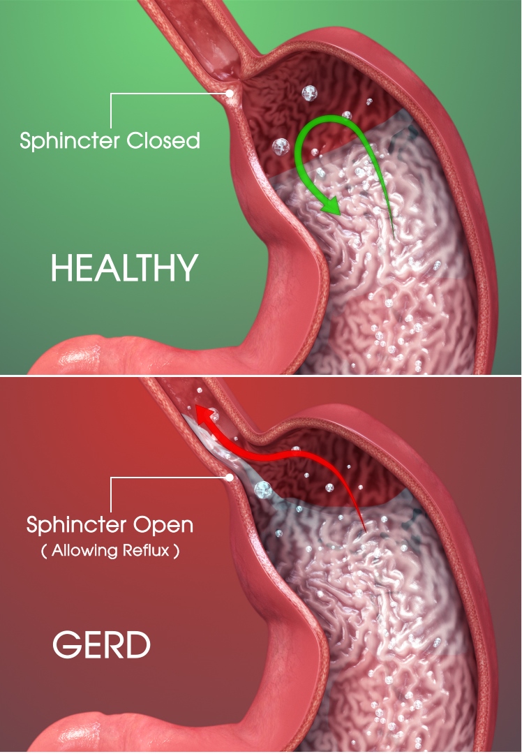
In acid reflux, the contents of the stomach make their way up into the esophagus and in some cases, even the throat and, believe it or not, the nose (this will cause a burning sensation).
Triggers of Acid Reflux
- This can be triggered by certain foods, particularly spicy.
- Heavy eating, particularly rushed, can also lead to it.
Dr. Zinberg further explains, “With each swallow, even subconsciously, air is swallowed as well (aerophagia), and this leads to burping of the accumulated air.
“Other causes of aerophagia include post-nasal dripping and anxiety.”
Preventing Burping from Acid Reflux
You’ll want to pay more attention to the kinds of foods that you eat. Acid reflux is commonly called heartburn because it causes a burning sensation in the chest.
But as Dr. Zinberg mentioned, it can also produce only one symptom: that of burping. And of course, belching in the presence of others is far more embarrassing than is feeling some heartburn.
So as far as the foods you should avoid or cut back on, they are as follows:
- Liquor, especially red wine
- Garlic, raw onions, black pepper
- Spicy foods
- Chocolate
- Caffeinated beverages
- Citrus fruits and citrus juices
- Peppermint
- Tomatoes
Another way to help prevent or reduce acid reflux is to avoid eating a lot of food at once.
In addition, if you tend to rush through your meals, this could lead to some burping. Slow down if you’re a fast eater.
Be mindful of whether or not you’re swallowing air with each spoonful, forkful or drink of your beverage of choice, even if it’s just water. Reducing acid reflux means reducing burping.
Lorra Garrick has been covering medical, fitness and cybersecurity topics for many years, having written thousands of articles for print magazines and websites, including as a ghostwriter. She’s also a former ACE-certified personal trainer.
.
Top image: Shutterstock/Nik Stock
Does Frequency of Heartburn Affect Risk of Esophageal Cancer?

Just how often must you have acid reflux from GERD (heartburn) to be at risk for esophageal cancer?
Esophageal cancer is one of the most frightening cancers to have, because its prognosis is poor in so many cases.
The disease is frequently diagnosed at an advanced stage because early symptoms can be vague or mistaken for less serious conditions.
Advanced stages of esophageal cancer often involve significant tumor growth or spread to other organs, making it harder to treat effectively.
19.2% – Number of people who are alive five years after a diagnosis of esophageal cancer, according to the National Cancer Institute Surveillance, Epidemiology and End Results Program.
As mentioned, this disease is typically discovered only after it has spread, making successful treatment very unlikely for the long-term.
There’s over 22,000 cases of new esophageal cancer diagnoses each year in the U.S., says the American Cancer Society.
People age 20 to 44 make up 2.3% of the cases, while those 65 to 74 make up 30.6%.
Heartburn, Acid Reflux
You may already know that GERD or chronic heartburn can lead to Barrett’s esophagus, which in turn is a risk factor for cancer of the esophagus.
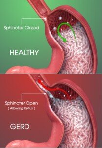
Scientific Animations, Creative Commons/BY-SA/Attribution-ShareAlike 4.0 International
“Generally, we consider high risk for Barrett’s esophagus and subsequent esophageal cancer, those who have prolonged reflux symptoms requiring medication at least three times per week for more than five years, especially if older than 50,” explains Jonathan Zinberg, MD, chief of gastroenterology at South Nassau Communities Hospital, Oceanside, NY.
“However, new onset symptoms past the age of 50 should also be suspect and evaluated.
“Some patients with Barrett’s have few if any symptoms, so these are merely guidelines for higher suspicion.
“Those with a family history of esophageal cancer are also at a somewhat higher risk.”
If you have GERD, commonly known as acid reflux, you need to prevent Barrett’s esophagus.
Barrett’s esophagus is a condition where the lining of the esophagus changes due to chronic acid exposure, increasing the risk of esophageal cancer.
To control GERD, consider making dietary changes, such as avoiding spicy, fatty or acidic foods, and refrain from eating close to bedtime.
Elevating your chest (not just the head) while sleeping can also help reduce acid reflux by keeping stomach acids from traveling up the esophagus.
If Barrett’s esophagus is diagnosed, lifelong surveillance is necessary.
This involves regular endoscopic examinations to monitor the esophagus for any precancerous changes or abnormalities.
Don’t be afraid of an upper endoscopy. This procedure is a valuable diagnostic tool that allows doctors to examine the esophagus, stomach and the beginning of the small intestine.
An upper endoscopy is typically performed using a thin, flexible tube with a camera, called an endoscope, which is gently inserted through the mouth and into the digestive tract.
The procedure is done under sedation, so you won’t feel discomfort during the examination.
In fact, you may even completely fall asleep from the sedation during the procedure, and after what seems like only a minute or two, you’re told that the procedure is over.
If any abnormalities are found, the doctor can often take biopsies for further analysis or provide immediate treatment.
Lorra Garrick has been covering medical, fitness and cybersecurity topics for many years, having written thousands of articles for print magazines and websites, including as a ghostwriter. She’s also a former ACE-certified personal trainer.
Source: seer.cancer.gov/statfacts/html/esoph.html

How Stress & Anxiety Cause Acid Reflux

Learn about mechanisms behind stress or anxiety causing acid reflux.
You may know full-well that acid reflux can be caused or triggered by anxiety and stress. But just how does this work?
“Stress may cause increased vagal nerve stimulation in the brain, which may lead to increased acid production in the stomach,” says Jonathan Zinberg, MD, chief of gastroenterology at South Nassau Communities Hospital, Oceanside, NY.
This was his first thought regarding this under-researched phenomenon.
Dr. Zinberg then explains, “There are many theories regarding stress and heartburn. Some say that increased cortisol/epinephrine leads to less blood flow to the stomach, with slow digestion and emptying of the stomach, with subsequent reflux.”
He continues, “Some point to decreased prostaglandin [a hormone] in the stomach, which helps to protect the lining of the stomach (so what?).
“Others blame stress for causing general poor habits (drinking alcohol, eating fatty foods, late bingeing and smoking) for causing worse reflux (maybe in some).”
Research Lacking
“There is little evidence for any of this,” says Dr. Zinberg. “The theory I mentioned about the vagus nerve sounded good, but I found no evidence for that either [after digging further into this after I sent in a few follow-up questions].
“In fact, a scientific study from 2005 showed no evidence for increased acid reflux at all during stress, despite increased symptoms of heartburn.
“So stress probably causes an increased perception of pain from minor stimuli rather than actual increased reflux.”
This may very well apply to some individuals, while others will swear that stress or anxiety (acute or chronic) is strongly associated with symptoms that clearly spell acid reflux.
I myself once developed what could only be acid reflux making its way into my throat and causing a cough every few minutes.
It’s no coincidence that at the time the cough developed, I was facing a colonoscopy to find out why I had sudden-onset diarrhea and weird-looking stools (I feared colon cancer).
And the procedure was two days after my beloved dog was euthanized for brain cancer — after I spent eight agonizing weeks trying to save him.
If this isn’t severe chronic anxiety and stress, I don’t know what is.
The cough vanished immediately after the gastroenterologist told me that everything looked normal, and it never returned.
I also developed a mild burning in my upper abdomen, plus dull aching in my chest, that coincided perfectly with the onset of a urine and feces stench that began invading my home through the ventilation and pipes — from the hoarder neighbor next door who was literally living in his wastes and accumulated garbage.
I was told by several authorities that nothing could be done about this.
I was faced with living with this disgusting foul odor in my own home indefinitely.
I was on the phone for hours on end with every conceivable authority in an attempt to get the neighbor evicted. If this isn’t extreme anxiety and stress, I don’t know what is.
The acid reflux symptoms vanished (and never returned) the day the police chief contacted me with news that the landlord was served a notice to remove the tenant within 72 hours!
How do stress and anxiety cause acid reflux? Maybe the best answer is: You decide.
Lorra Garrick has been covering medical, fitness and cybersecurity topics for many years, having written thousands of articles for print magazines and websites, including as a ghostwriter. She’s also a former ACE-certified personal trainer.
.
Top image: Freepik

Cause of White Sticky Slime in Mouth Walls & Prevention

Finally, an explanation for what causes that gooey, white, slimy substance inside the walls of your mouth.
As annoying as that white, sticky and gooey substance inside the walls of your mouth is, it’s actually not a medical issue and nothing to worry about.
Yes, it’s gross, and it’s embarassing when it makes its way to the corners of your lips and someone asks you about it.
But other than inconvenience and potential embarassment around other people, that gooey stuff is actually nothing to worry about in terms of your health.
What causes the white, sticky substance inside the mouth?
“Oral mucosa, like skin cells, will slough off as newer mucosal or skin cells develop,” explains Dr. Stacey Silvers, MD, of Madison ENT & Facial Plastic Surgery in NYC, who is board certified in otolaryngology.
“This can be exacerbated by certain mouthwashes and toothpastes.
“Especially if the toothpaste or mouthwash is in for an excessive period of time. This is similar to exfoliating the skin.”
If you’ve had the problem of a white, slimy and sticky goop inside your mouth, have you noticed it forms after you’ve brushed your teeth or used a mouthwash?
Your Toothpaste Could Be Causing a Sticky White Slime
Toothpaste can sometimes cause a white, slimy substance to form inside the mouth, which may be due to a few different factors.
1. Sodium Lauryl Sulfate (SLS): Many toothpastes contain sodium lauryl sulfate, a common foaming agent.
For some people, SLS can cause irritation or a reaction in the mouth’s soft tissues, leading to the formation of a white, mucous-like substance.
This reaction can result in a slimy or coating effect inside the mouth. Avoid toothpastes that contain this chemical.
2. Oral Mucosa Reaction: The mouth’s mucous membranes might react to certain ingredients in the toothpaste, such as flavorings, preservatives or fake colorings.
3. Dry Mouth: Toothpaste can sometimes contribute to dryness in the mouth, particularly if it contains alcohol or other drying agents.
Dry mouth can lead to an accumulation of dead cells and bacteria, which may appear as a white film.
4. Bacterial Growth: A white, slimy goo could also indicate an overgrowth of bacteria or yeast in the mouth, which might be aggravated by certain toothpaste brands ingredients.
Switch toothpaste brands and see what happens. Not all toothpastes are the same. The one that finally works best for you may be pricier, but the extra cost will be well worth it.
If this article provided you with helpful information or reassurance, please consider supporting my work through Ko-fi. Donations help me continue writing detailed medical content that’s easy to understand.
 An NYC expert in ear, nose and throat care, Dr. Silvers has been named among America’s Top Physicians and Surgeons in facial plastic surgery and otolaryngology numerous times since 2003. Dr. Silvers is an expert in the field of minimally invasive rhinology, resolving patients’ breathing and sinus problems with simple in-office procedures.
Lorra Garrick has been covering medical, fitness and cybersecurity topics for many years, having written thousands of articles for print magazines and websites, including as a ghostwriter. She’s also a former ACE-certified personal trainer.
.
Top image: Shutterstock/Alexander Raths








Under-Eye Wrinkles: Have Surgery; TCA Peel Won’t Work

A TCA peel won’t work for under-eye wrinkles, says a plastic surgeon.
A big question out there is:
Does a TCA peel work for wrinkles under the eyes?
“TCA is an excellent peel, but it doesn’t work for under-eye wrinkles,” says Allen Gabriel, MD, board certified plastic surgeon with PeaceHealth Medical Group Plastic Surgery in Vancouver, WA.
“This is usually caused by thinning of the skin and weakening of the under-eye muscle, or even the bulging of under-eye fat, all of which can be improved surgically.”
How does a TCA peel work?
Dr. Gabriel explains, “TCA is a great way of rejuvenating the skin. It works by removing the top layers of the skin and after the skin sloughs (3-7 days), the new rejuvenated skin is exposed.
” This helps with pigmentations and melasma and can overtime support with regenerating collagen as long as other prescription products are used in conjunction such as retin-A.
“However, under-eye wrinkles are multifactorial and generally a surgical treatment is needed to correct the deformities already stated. Combination of skin treatments and surgery is ideal, as it will add to the longevity of the results.”
How to Minimize Under-Eye Wrinkles in the First Place
You should be very cognizant of what your hands and fingers do when they make contact with your face.

Freepik.com
Do you ever rub or scratch at the skin below your eye? This pulling action, when done too much and over time, can loosen the skin.
What about how you clean the skin below your eye? Does this involve any pulling, e.g., rubbing at the area with a Q-tip swab in an attempt to remove dirt or makeup?
What about when you’re at the computer with your face in a palm? This, too, can elongate the skin.
Be alert to how you sleep. Sleeping on your side can stretch the delicate skin underneath your eye!
Finally, be very gentle and patient when you apply creams in the skin under the eye, such as sunscreen or moisturizer.
Don’t rub it in. Dab it on with the pad of a fingertip.
 Dr. Gabriel is adept at addressing a wide range of concerns, from breast reconstruction after mastectomy and the treatment of congenital anomalies, to complex facial and breast surgeries performed solely for cosmetic improvement.
Lorra Garrick has been covering medical, fitness and cybersecurity topics for many years, having written thousands of articles for print magazines and websites, including as a ghostwriter. She’s also a former ACE-certified personal trainer.
.