
Can a Skin Injury Turn into Cancer? Yes, but What Kind?
Yes, a skin injury can indeed develop into cancer, says a dermatologist.
There are three main types of skin cancer: melanoma, squamous cell carcinoma and basal cell carcinoma.
Can an injury to the skin lead to cancer?
“The answer is a resounding yes,” says Dr. Rebecca Tung, MD, a
“A past injury to the skin such as trauma from an accident which formed a scar, a vaccination site scar or even past exposure to radiation or caustic chemicals which resulted in scar formation can potentially turn into a skin cancer over time.”
How long does it take for a scar to morph into cancer?
“Typical malignant change may occur over decades in these cases,” says Dr. Tung.
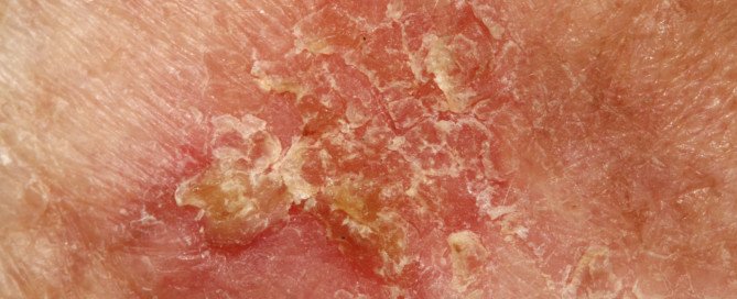
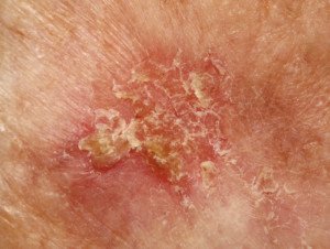
“Signs that may alert you that your scar is no longer ‘just a scar’ include pain, redness, bleeding or skin breakdown.
“Scars and skin which has been persistently inflamed can turn into a type of skin cancer called squamous cell carcinoma.”
What is squamous cell carcinoma?
“This type of SCC is rare and was initially coined as a ‘Marjolin’s ulcer’ after the surgeon who first described the condition.
“There have also been reports of the more common type of skin cancer called basal cell carcinoma occurring in vaccination scars. Burn scars can also be the site of skin cancer years after the burn has healed.”
Excessive Sun not Always a Factor
“While most non-melanoma skin cancers (BCCs and SCCs) result from sun or ultraviolet exposure, research shows that long-term inflammation and scar formation can also create cancerous change in the skin,” says Dr. Tung.
“Your dermatologist can confirm the diagnosis of skin cancer by performing a skin biopsy.
“A scar (increased density of collagen in the lower part of the skin called the dermis) looks a world of difference away from a skin cancer (collections or islands of atypical cells) under the microscope.”
Be on Lookout for Changing Scar
“Have a changing scar? You owe to yourself to see a dermatologist to help determine if it has transformed into cancer.
“Again, when skin cancer is detected in its early stages, it’s often fully curable with surgical removal.”
Dr. Tung’s specialties include general dermatology with skin cancer surveillance, moles, melanoma, surgery (Mohs micrographic, laser, skin cancer reconstruction) and cosmetic dermatology.
 Lorra Garrick has been covering medical, fitness and cybersecurity topics for many years, having written thousands of articles for print magazines and websites, including as a ghostwriter. She’s also a former ACE-certified personal trainer.
.
Top image: Shutterstock/Dermatology11

Stomach Cramps After Your Period Ends: Five Causes

Could abdominal cramping after menstruation ends be related to early cancer?
There’s actually a variety of causes for this troubling condition that all women should be aware of.
When a woman who’s completed her monthly cycle then has unexplained abdominal cramps — especially in the lower area of the abdomen — she might start fearing that the cause is ovarian or uterine cancer.
After all, these cancers DO strike fertile women. According to the National Cancer Institute Surveillance and Epidemiology and End Results Program, 28.9 percent of ovarian cancer diagnoses in the U.S. occur in women between 20 and 54. For uterine cancer it’s 23.4 percent.
What benign condition can cause abdominal cramping after menstruation ends?
This depends on how long after the period ends, that a woman experiences abdominal cramping, says Wendie Trubow, MD, an OB/GYN with Five Journeys, a membership-based wellness organization that uses functional and integrative medicine for evaluation and treatment.
In a menstrual cycle that lasts seven days, ovulation for the next cycle may begin in just a few days.
“So the cramping can be due to release of a dominant follicle from one of the ovaries,” says Dr. Trubow.
“This typically causes some fluid in the pelvis, and that can cause irritation and cramping.”
In a typical menstrual cycle, ovulation usually occurs around the middle of the cycle, often about 14 days before the start of the next period.
However, if your cycle lasts only seven days, this suggests a shorter overall cycle.
For example, if your cycle lasts 21 days, ovulation could happen around day 7, which means you may be close to ovulating as soon as the current cycle ends.
In this case, ovulation could potentially happen just a few days after your period ends.
Other Causes of Stomach Cramping Following a Period Include Cancer
“The cramping could also be due to fibroids, or a polyp or an IUD (intrauterine device),” says Dr. Trubow.
Discomfort from any of these conditions “is due to the body trying to get rid of a foreign body, and the uterus only really knows how to do a few things, including grow to accommodate a fetus, and contract, to get rid of whatever doesn’t need to, or shouldn’t be there.”
Irritable bowel syndrome and microscopic colitis (both are inflammatory bowel diseases) can cause cramping in the stomach or pelvic region.
Both conditions, though potentially capable of impacting one’s quality of life, are benign and can’t lead to cancer.
But speaking of cancer, ovarian cancer and uterine cancer can cause an abdominal cramp as well.
 Having practiced functional medicine since 2009, Dr. Trubowworks with women to identify causes of their symptoms and then works with them to achieve their greatest health and life balance.
 Lorra Garrick has been covering medical, fitness and cybersecurity topics for many years, having written thousands of articles for print magazines and websites, including as a ghostwriter. She’s also a former ACE-certified personal trainer.
Top image: Shutterstock/Dragana Gordic
Source: seer.cancer.gov

Why Does Morbid Obesity Stop Menstruation?

Being morbidly obese, no matter how confident you are, can have a bad effect on your menstrual cycle.
It is no secret that when a very obese woman has been unable to conceive despite a lengthy time period of attempts, her size is to blame when all other tests — including those of her husband — have turned out normal.
“Morbid obesity can have either effect; either to stop menstruation, or to increase it,” says Wendie Trubow, MD, an OB/GYN with Five Journeys, a membership-based wellness organization that uses functional and integrative medicine for evaluation and treatment.
How Morbid Obesity Influences a Woman’s Period
“The mechanism is this: Circulating androgens are converted into estrogen by fat cells,” begins Dr. Trubow.
The more fat that you have in your fat cells, “the more conversion to estrogen. Ovulation occurs by ‘spikes’ in estrogen and progesterone.
“A constant stream of estrogen inhibits ovulation. If a woman does not ovulate, but has circulating estrogen, which serves to build the uterine lining, it will eventually shed.
“However, since the woman didn’t ovulate, the shedding doesn’t occur in an organized fashion (which is a typical menstrual flow).
“It occurs in a disorganized fashion, which can occur in varied amounts, at any time.”
So while some morbidly obese women struggle with reduced or ceased menstruation, others experience an increased flow of blood — which is never a picnic.
Solution to Menstrual Issues Caused by Obesity
The solution is weight loss. If you desperately are trying to conceive, you may want to put that plan on the shelf for awhile as you pursue safe weight loss via healthier eating, portion control, aerobic exercise and intense strength training.
Do not go on a crash diet to get the job done faster out of eagerness to conceive.
If you have the opposite problem — heavy periods — again, see if safe weight loss doesn’t correct this problem over time.
Having practiced functional medicine since 2009, Dr. Trubow works with women to identify causes of their symptoms and then works with them to achieve their greatest health and life balance.
Lorra Garrick has been covering medical, fitness and cybersecurity topics for many years, having written thousands of articles for print magazines and websites, including as a ghostwriter. She’s also a former ACE-certified personal trainer.

Causes of Slow, Prolonged Menstrual Bleeding

Find out what can cause bleeding from your period to be slow and prolonged.
“Perhaps the most common cause of this is when the blood has difficulty passing through the cervical canal, due either to obstruction (perhaps a fibroid is in the canal), or to the cervix simply being ‘tight,’” says Wendie Trubow, MD, an OB/GYN with Five Journeys, a membership-based wellness organization that uses functional and integrative medicine for evaluation and treatment.
What can cause a tight cervix?
Dr. Trubow explains that this can be due to not yet having given birth, “or if the woman has had surgery on the cervix to remove abnormal cells and had scarring in the cervix, which makes it harder for the cervix to open to let the blood through.”
Another Cause of Slow, Prolonged Menstruation
“Alternatively, the bleeding could be slow due to inability of the uterus to contract, which can usually be attributed to fibroids,” says Dr. Trubow.
“Lastly, this could be bleeding due to an anovulatory [absent] cycle, so the bleeding is simply occurring in a disorganized fashion.”
Cancer and Vaginal Bleeding
Bleeding in between periods, or vaginal bleeding after menopause, needs to be checked out by a gynecologist because uterine, cervical and vaginal cancer can cause this symptom.
However, if you know the behavior of your period very well, and can recognize that the slow or prolonged bleeding is part of your menstruation, then the chances of this situation being caused by cancer are not likely.
Nevertheless, any kind of bleeding or spotting that is out of the ordinary, that has no confirmed explanation, needs to be evaluated by a doctor.
You should keep a documentation of the nature of your monthly cycles so that you can get acquainted with what your normal is.
Some women do spot before and after four or five days of actual flowing.
Having practiced functional medicine since 2009, Dr. Trubowworks with women to identify causes of their symptoms and then works with them to achieve their greatest health and life balance.
Lorra Garrick has been covering medical, fitness and cybersecurity topics for many years, having written thousands of articles for print magazines and websites, including as a ghostwriter. She’s also a former ACE-certified personal trainer.
.
Top image: Shutterstock/Evan Lorne
How Common Is a Flesh Colored Melanoma?
Here’s what a dermatologist has to say about frequency of flesh colored melanoma — which can go undetected during your self-exams.
Melanoma can present as flesh colored.
But just how often does this occur?
How common are flesh colored melanomas?
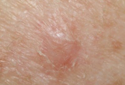
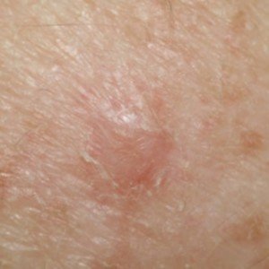
“Amelanotic (flesh colored) melanomas are a rarer type of melanoma,” says Dr. Rebecca Tung, MD, a
“The incidence of amelanotic melanoma is only 2-8 % of all melanomas.
“They often appear as flesh colored to reddish lesions, often on the skin of the trunk, and do not follow the usual ABCD rules (asymmetry, border irregularity, color changes, diameter of 6mm or larger or growing in size) of melanoma detection.”
Sometimes, this flesh or pinkish/red colored melanoma can pass as basal cell carcinoma or squamous cell carcinoma.
A basal cell carcinoma is the most common cancer in the world. This easily curable tumor grows very slowly and very rarely spreads beyond locally.
Squamous cell carcinoma can spread, but it’s nowhere near as deadly as melanoma.
Both of these skin cancers are strongly linked to exposure to the sun.
Alarmingly, the amelanotic melanoma can also pass as a common pimple or blemish.
“Patients often initially mistake this type of melanoma as scars or pink moles,” continues Dr. Tung.
“Even under a special magnifying device called a dermatoscope, the features which usually signal cancer are often absent.
“For these reasons, diagnosis is frequently delayed.”
If you don’t have a good feeling about a new spot and especially “pimple” on your skin, you should have it removed.
“Suspicious lesions should be biopsied to confirm the diagnosis,” says Dr. Tung.
“Treatment is surgical with wide local excision.
“If the lesion extends deeply into the skin, lymph nodes may also need to be sampled (a procedure called sentinel lymph node biopsy) to determine if they do or do not contain melanoma.”
Dr. Tung’s specialties include general dermatology with skin cancer surveillance, moles, melanoma, surgery (Mohs micrographic, laser, skin cancer reconstruction) and cosmetic dermatology.
Lorra Garrick has been covering medical, fitness and cybersecurity topics for many years, having written thousands of articles for print magazines and websites, including as a ghostwriter. She’s also a former ACE-certified personal trainer.

Causes of White Peeling Skin Inside Lip Include Cancer

A dermatologist names cancer as a possible cause of white peeling skin inside the lip.
In addition to having just brushed one’s teeth, which can cause cells inside the lip to slough off, other conditions can cause the inside of the lip to develop a white, peeling reaction.
“Lichen planus (LP) is a condition that can affect the mucus membranes of the mouth,” says Dr. Rebecca Tung, MD, a
“It often looks lacy in a lattice pattern. While most cases occur on their own as a result of a body’s altered immune response, other cases can be the result of sensitivity or allergy to metal in dental work, or even a skin finding of an infectious viral hepatitis (inflammation of the liver; hepatitis C).”
Cancer As Cause of White Peeling Inside the Lip
“A more serious white patch on the inside of the bottom lip could be skin cancer — specifically squamous cell carcinoma (SCC) or its precancerous state (leukoplakia),” says Dr. Tung.
“Smokers, tobacco chewers and heavy drinkers are at increased risk for SCC.
“A biopsy of the lesion can confirm the diagnosis and direct treatment.”
In fair skinned people, squamous cell carcinoma is the second most common skin cancer, after basal cell carcinoma.
SCC is highly curable when caught early, but it kills thousands of Americans every year.
Dr. Tung’s specialties include general dermatology with skin cancer surveillance, moles, melanoma, surgery (Mohs micrographic, laser, skin cancer reconstruction) and cosmetic dermatology.
Lorra Garrick has been covering medical, fitness and cybersecurity topics for many years, having written thousands of articles for print magazines and websites, including as a ghostwriter. She’s also a former ACE-certified personal trainer.
.
Top image: Shutterstock/Image Point Fr

Causes of Itchy, Flaky Skin on Your Face

Are you worried that cancer could be causing that unexplained itching and flaking on your face?
The causes of itchy, flaky facial skin are many.
Eczema. “Scaling on the face can be as simple as eczema (atopic dermatitis)—this occurs in children and adults when the barrier function of the skin is altered and the skin is hypersensitive,” says Dr. Rebecca Tung, MD, a
“Oftentimes it is very itchy and may even become infected in patients who aggressively scratch their skin.
“Minimizing the itch-scratch cycle is the cornerstone of treatment — oral antihistamines, topical or oral steroids, and topical medicines that reduce the hyperactive immune response.”
Contact dermatitis. “A specific type of dermatitis called contact dermatitis can cause similar symptoms,” says Dr. Tung.
“However, the culprit is some product or ingredient that is applied to the skin such as a fragrance in a facial cream, preservative in lotion, or even a particular antibacterial agent like neomycin in an over-the-counter wound healing ointment.”
Dr. Tung has even seen flaking on the skin from applying body glitter, which contains nickel, the allergen.
“Even some types of chemical sunscreens can bring on scaly, red, itchy patches on the face. Finding the cause is critical to cure.
“This is accomplished with specialized allergy testing (patch testing)—no needles involved under the guidance of a dermatologist.”
Seborrheic dermatitis. Dr. Tung explains, “Other times infection with a specific type of yeast (Malassezia species) can bring on a case of facial redness and scaling, particularly around the eyebrows, nose and ears as well as is the scalp (dandruff).
“Treatment is successful when the yeast are killed (anti-fungals—topical or oral) and inflammation is minimized (topical steroid).
Others causes of itchy, flaking skin on the face.
“Sometimes if the redness and scaling is accompanied by sun sensitivity—facial redness can signify an autoimmune condition like lupus or dermatomyositis.
“Your dermatologist may order blood tests or perform a skin biopsy to confirm the diagnosis.”
Dr. Tung’s specialties include general dermatology with skin cancer surveillance, moles, melanoma, surgery (Mohs micrographic, laser, skin cancer reconstruction) and cosmetic dermatology.
Lorra Garrick has been covering medical, fitness and cybersecurity topics for many years, having written thousands of articles for print magazines and websites, including as a ghostwriter. She’s also a former ACE-certified personal trainer.
.
Top image: ©Lorra Garrick

Causes of Anal Lumps Include Cancer

A dermatologist says there are several causes of anal lumps, and one is cancer.
Most causes of anal lumps are not related to cancer, but cancer can be a cause.
“Lesions around the anus can be as harmless as hemorrhoids (except of course if these hemorrhoids are painful or bleeding—this requires surgical treatment with a colorectal surgeon),” says Dr. Rebecca Tung, MD, a
Skin tags can be another cause of anal lumps, though they won’t really look like true lumps, but they may feel like them against your fingertips. Skin tags are collections of extra tissue.
Other Benign Causes of Anal Lumps
Dr. Tung adds that a skin infection that causes genital warts can be a culprit.
Cancer
Squamous cell carcinoma is a type of skin cancer that can cause anal lumps, says Dr. Tung.
“Fortunately, appearances of each of these conditions are fairly specific, so a skin biopsy is only needed when the look is not typical.
“Skin tags can be numbed with a topical anesthetic and then snipped and cauterized.
“Skin cancers require surgical and possibly additional medical treatment.
“Warts can be treated with freezing (cryotherapy—liquid nitrogen application ), topical medicine application or other minimally invasive procedures like spot chemical peeling, electrical cautery or laser surgery.”
Dr. Tung’s specialties include general dermatology with skin cancer surveillance, moles, melanoma, surgery (Mohs micrographic, laser, skin cancer reconstruction) and cosmetic dermatology.
Lorra Garrick has been covering medical, fitness and cybersecurity topics for many years, having written thousands of articles for print magazines and websites, including as a ghostwriter. She’s also a former ACE-certified personal trainer.
.
Top image: Freepik

Can White Pimples on Penis Be Caused by Cancer?

A dermatologist names some causes of white bumps or pimple-like growths on the penis.
White pimples, bumps or small growths can appear on one’s penis; what are some causes?
“The most benign condition that causes flesh colored lesions on the penis is called pearly penile papules—not precancerous or caused by infection—just benign non-cancerous growths that can form a ring around the shaft,” says a
“Other flesh colored lesions could be genital warts (condyloma). Since they are caused by a virus, early treatment is a good idea to get rid of current lesions and prevent spread of new lesions.
“Other white bumps on the penis can be simple cysts (called milia)—harmless and do not require treatment unless bothersome.”
It is extremely unlikely that a white “pimple” or “bump” on the penis can be skin cancer or any kind of cancer.
Dr. Tung’s specialties include general dermatology with skin cancer surveillance, moles, melanoma, surgery (Mohs micrographic, laser, skin cancer reconstruction) and cosmetic dermatology.
Lorra Garrick has been covering medical, fitness and cybersecurity topics for many years, having written thousands of articles for print magazines and websites, including as a ghostwriter. She’s also a former ACE-certified personal trainer.
.
Top image: Shutterstock/sheff

Can a Red Spot on the Lip Be Cancer?

A dermatologist lists many causes of a red spot the lip; find out if cancer is one.
What can cause a red spot on the lip?
Can cancer be a cause?
Rebecca Tung, MD, explains that often, a red spot(s) on one’s lip can be “small collections of blood vessels going by names such as telangiectasia, angioma or venous lake.”
These are benign and can also appear elsewhere on the body.
“Most often there is no particular cause and can be easily treated with a vascular laser,” continues Dr. Tung, a
More Serious Causes of a Red Spot on the Lip
“However, if there are sprays of blood vessels (broken capillaries) on the lips and face/body — this may signal a more serious condition called hereditary hemorrhagic telangiectasia,” says Dr. Tung.
“Affected people are at risk for nosebleeds, acute bleeding in the gut and other internal organs.
“Treatments circles around reducing bleeding from blood vessel lesions.
“Another condition where you can see widespread vascular lesions and thickening of the skin is called scleroderma.
“A particular type of this autoimmune disease is called CREST syndrome—calcinosis (calcium deposits in the skin).”
If you have scleroderma, you’ll have other symptoms far more troubling than a red spot on your lip.
Another condition that can cause red spots on the lip is Raynaud’s syndrome, a vascular disease that affects the legs and other parts of the body.
Dr. Tung names more conditions: “Esophageal dysmotility (cannot swallow properly), sclerodactyly (finger skin becomes abnormally thickened and easily forms ulcers—very painful).
“Fortunately, medicines which adjust the body’s immune response are often helpful.”
As you can see, cancer did not make the list of conditions that can cause a red spot to appear on the lip.
However, if you’re still concerned, ask a dermatologist to inspect it with a dermatoscope, a handheld lighted lens.
Dr. Tung’s specialties include general dermatology with skin cancer surveillance, moles, melanoma, surgery (Mohs micrographic, laser, skin cancer reconstruction) and cosmetic dermatology.
Lorra Garrick has been covering medical, fitness and cybersecurity topics for many years, having written thousands of articles for print magazines and websites, including as a ghostwriter. She’s also a former ACE-certified personal trainer.


