
What exactly is a dermatoscope? First off, next time you see a dermatologist for a skin cancer screening, make sure the doctor is using a dermatoscope.
Sometimes, a dermatoscope is incorrectly called a dermoscope. “Derma” means skin, and “scope” means to view.
A dermatologist, when examining a patient’s moles for signs of melanoma, should use a dermatoscope.
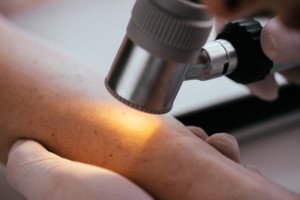
“This is a specialized dermatologist’s tool that uses polarized light and magnification to spot irregularities in the skin,” says Bobby Buka, MD, a NYC dermatologist and CEO of The Dermatology Specialists, and author of “Buka’s Emergencies in Dermatology” and “Top 50 Dermatology Case Studies for Primary Care.”
“It’s a handheld device that your doctor can use to inspect spots wherever they occur,” continues Dr. Buka.
“The polarization of the light it emits allows us to more clearly see pigment inconsistencies that can indicate melanomas or other skin problems.”
A more sophisticated model has several magnifying lenses together that are in a tube with a polarized or non-polarized light source.
The more complex the instrument, the more information a doctor will get when viewing your moles and other skin lesions.
For instance, a seborrheic keratosis may look very much like a common mole – until the doctor views the former through a dermatoscope.
The newest instruments have a bright LED light encircling the magnifying lens. There may also be a digital camera, which is very useful when a mole wants to be tracked over time.
To get a close-up inspection of the skin, a doctor will place the dermatoscope against the skin and look very close into it.
The tool will likely have liquid that’s between the lens and the skin to reduce light glare and scatter, improving visibility of the lesion.
When a normal mole is viewed through this device, it can look “bad” to the layperson, because the layperson doesn’t know what’s normal or what’s suspicious.
This is why laypeople should not purchase these devices, which are sold online.
A layperson with health anxiety will set himself up for much anxiety by getting into the habit of inspecting their moles with a dermatoscope.
Dermatologists spend enormous hours of training and learning what characteristics in a mole to look for that are suspicious for melanoma.
After looking at thousands of different kinds of skin lesions that can pass for melanoma, as well as actual melanomas, doctors will still be crafting their expertise at knowing what they’re looking at.
Can a Dermatoscope Diagnose Melanoma?
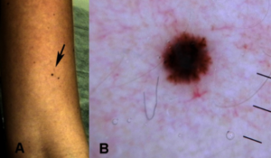
Dermatol Pract Concept. 2013 Apr Copyright ©2013 Pellizzari et al
There’s no doubt that this tool can show classic features of melanoma.
But it is not a diagnostic tool.
A doctor will inform the patient that the lesion should be biopsied.
Not all the biopsy results, however, come back positive for disease.
A mole can look suspicious under the magnifying lens but come back benign.
Other Uses of a Dermatoscope
• Evaluation of skin capillaries
• Assess patterns of hair follicles
• Examine other conditions like actinic keratosis
Remember, don’t go buying a dermatoscope to check your moles! You don’t know what to look for. Leave it up to your dermatologist.
 Dr. Buka is the contributing Founder and Chief Science Officer of the First Aid Beauty skin care line. firstaidbeauty.com
 Lorra Garrick has been covering medical, fitness and cybersecurity topics for many years, having written thousands of articles for print magazines and websites, including as a ghostwriter. She’s also a former ACE-certified personal trainer.
.


