

Teen Girl Is Overweight, Overeats: Is this an Eating Disorder?

Overeating in a nation that makes it easy for teens to overeat — and be overweight — doesn’t always mean an eating disorder.
Now keep in mind that in a culture where fast-food restaurants, bakeries, donut shops, bagel shops and burrito stands are there at every turn, it’s easy to overeat junk food. It’s THERE.
You can smell the aroma wafting through the air at the mall. You follow the aroma of the cinnamon rolls or giant soft pretzels.
And it all just tastes SO good!
But there comes a point where the situation may go beyond filling a hungry stomach with fabulous tasting foods.
The eating continues well-past satiation, and is triggered by circumstances such as boredom, stress, anxiety and depression.
Weight Loss through Exercise for Overweight Teen Girls
Exercise is good for every body. But like eating, exercise should never be done mindlessly. It should have a tactical approach.
The No. 1 reason for exercising should be for fitness and health. But if you want to lose weight, it’s perfectly okay to have fat loss in mind when working out.
Nevertheless, two hours jogging around the neighborhood in the name of losing weight is NOT the answer.
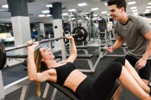
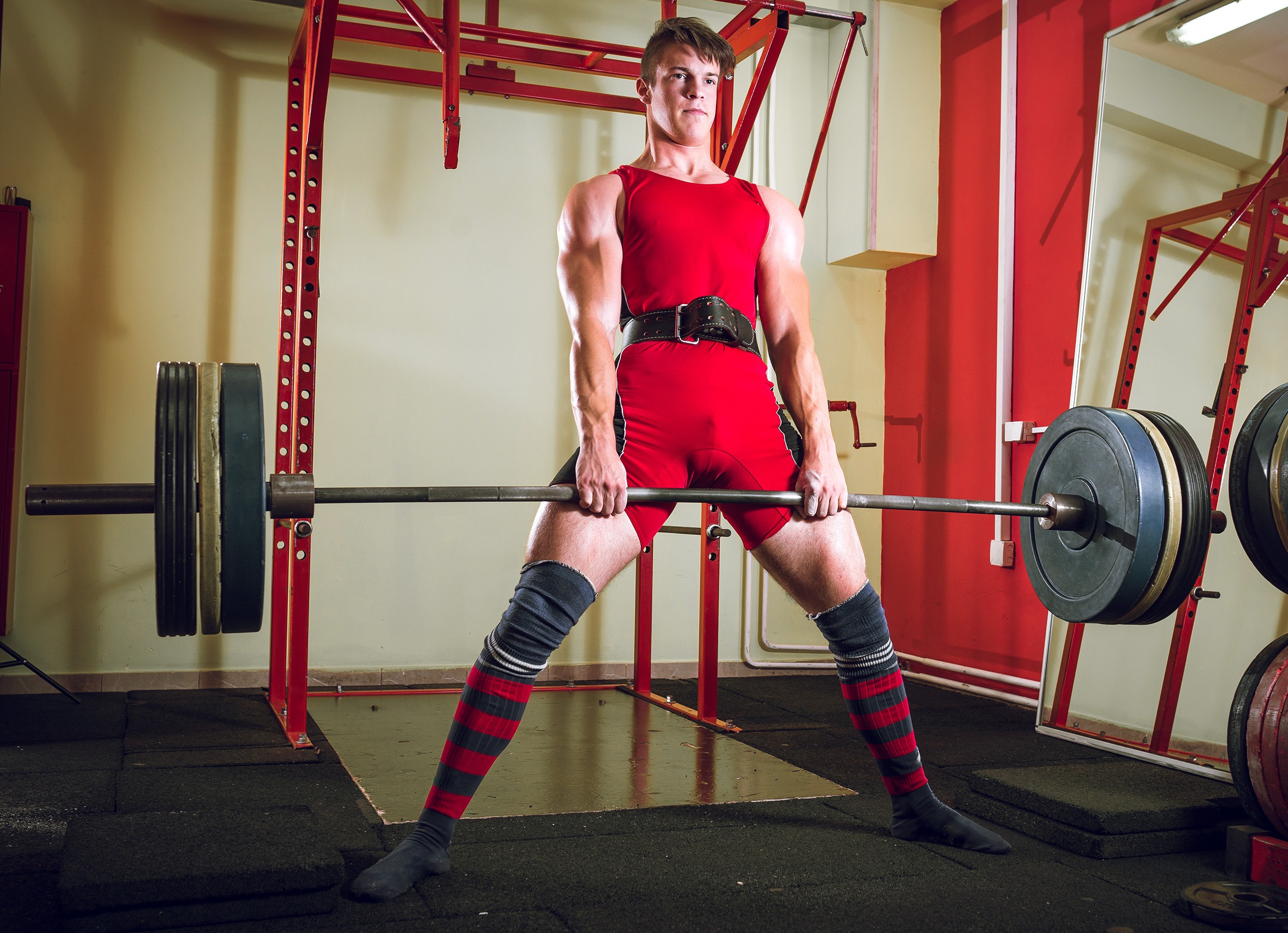
Strength Training
A teen girl is not too young to start lifting weights. And you are never “too fat” to lift weights. And lifting weights will NOT make you bigger!
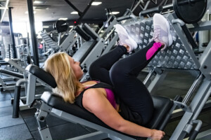
Shutterstock/Aleksey Boyko
Which might make you bigger? Picking up dumbbells or picking up donuts?
Never believe for a second that working out with weights will increase your size.
Just remember this: If you feel bad about your body, it’ll be rather difficult to continue feeling this way once you’ve trained your body to lift heavy things and find that you can shovel snow or haul out the garbage effortlessly.
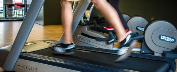
Aerobics
As a former personal trainer, I recommend high intensity interval training. This solves the problem of time investment and minimizes obsession.
Just 20 minutes of HIIT will burn more fat than two hours of slow paced cardio.
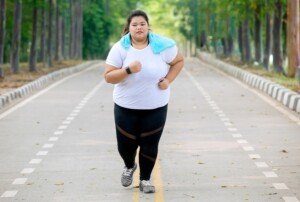
Shutterstock/Creativa Images
Plus, since HIIT can be done in only 20 minutes, after which you’ll feel spent, you’ll less likely become obsessed with it.
Whereas paced cardio can be done for three hours straight – making it easier to become obsessed with. Here are instructions on how to do HIIT.
Overweight in Teens: Don’t Wait till It Gets Worse
Addressing weight concerns now does not mean a breach of body positivity.
Rather, it’s about preventing type 2 diabetes, high blood pressure and other obesity related issues (e.g., difficulty conceiving) down the road during adulthood.
As a teen, you can still feel positive about your body (which is easier to do when you can lift heavy barbells) while working on losing surplus weight in a safe and realistic way.
 Lorra Garrick is a former personal trainer certified through the American Council on Exercise. At Bally Total Fitness she trained women and men of all ages for fat loss, muscle building, fitness and improved health.
.
Top image: Freepick.com/brgfx

Women’s Strength Training Rules for Maximum Fat Loss

If you’re disgusted with the fat in your thighs, tummy or elsewhere that just won’t budge, it’s time to take control with a strength training routine that will blast off the fat.
I’m a former certified personal trainer and will describe in detail how a woman can use strength training to achieve the greatest amount of fat loss anywhere in her body.
In order for strength training to burn maximal fat, strength training must be done very intensely.
There is a correlation between intensity and how heavy the weight is. However, if the weight is too heavy, you won’t get the intensity. Intensity is achieved with repetitions.
So if the weight is so heavy that you can lift it only five times, this won’t be enough reps to induce “metabolic failure,” which is what you want.
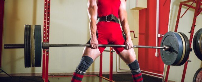
For optimal fat loss, a woman should do strength training with an amount that brings muscle failure (the metabolic type) within 8-12 reps.
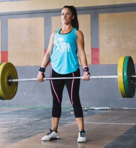
Deadlift. Freepik.com
Many women already do 8-12 reps for their strength training – simply because they’ve read or heard that this is the ideal rep range.
Try this experiment: Next time you do your strength training, use the same amount of weight you normally use, but instead of stopping at between 8 and 12 reps, see if you can go to 15. If you can reach 15, see if you can go to 18, even 20.
If you can do more than 12 reps, the weight isn’t heavy enough. So, if you can actually make it to near 20 reps, the weight is entirely too light.
For strength training to induce optimal fat burning, a woman must use a weight that she cannot lift more than 12 times, but at least 8 times.
This is called the 8-12 rep max (or RM). If you can lift the weight 13 times but not 14, it is still too heavy. Increase it a little so that you are forced to stop at 8, 9, 10, 11 or 12 reps.
Metabolic burn isn’t pretty. It hurts, but it’s a good kind of hurt. The weight can no longer be lifted because your muscles are burning so much, rather than the weight is too heavy, as in “mechanical failure” that would result from a 4-6 RM.
A woman’s rest time in between strength training sets for optimum fat loss

Bent-over barbell row. embhoo/CreativeCommons
Rest time is 45-60 seconds. This means that for many sets, you’ll need to lighten the weight to achieve an 8-12 RM.
For example, in a bench press, suppose your first set of 8-12 RM is at 65 pounds. After 45-60 seconds, you can only do 6 reps with the 65 pounds.
This means that for the third set, the weight should be lowered to 55 or maybe 50 pounds. If you can push 50 pounds 14 times, this is a little too light.
Strength training this way will require some experimentation of weight loads, obviously. But this is what successful fitness enthusiasts do all the time; it goes with the territory.
Which muscle groups should be strength trained for the most fat loss?
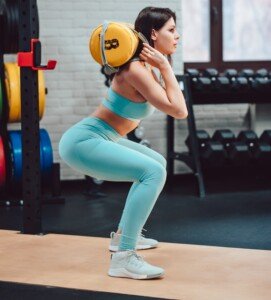
Half squat. Freepik.com, teksomolika
One 45 minute session a week should be devoted entirely to legs. Legs can be major fat-burners if the strength training is done right, because they are such large muscle groups.
On a second day of the week, do a 60 minute strength training session targeting the back and shoulders.
On a third day for 60 minutes, strength training the chest, triceps and biceps will do.
Incline press. Freepik.com, javi_indy
Should women strength train with mostly machines or free weights?
Most of the routines should be with free weights, though the following machines are great: lat pull-down, chest press, leg press, leg extension and leg curl.
So though seated chest press is fine, do some chest presses with a barbell or dumbbell also.
Number of sets per muscle group is variable; general rule is that the more muscles worked in a given exercise, the more sets for that muscle group.
This means that on leg day, do a lot of squats, leg presses and weighted lunges, but fewer leg extensions and leg curls. Do only a FEW sets of inner and outer thigh routines.
To burn the most fat, women should do heavy strength training and follow these guidelines, but ease gradually into this type of strength training to avoid injury or excessive soreness.
Lorra Garrick is a former personal trainer certified through the American Council on Exercise. At Bally Total Fitness she trained women and men of all ages for fat loss, muscle building, fitness and improved health.
.
Top image: ©Lorra Garrick
Why People Drive to the Gym to Use a Treadmill
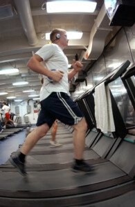
There’s a lot of logic behind driving to the gym to use a treadmill.
I just can’t believe that anyone would ask why a person would drive to a gym (instead of walk) and then walk on a treadmill.
But there’s actually a lot of rhyme and reason to doing this.
I’m a former certified personal trainer, and I’ve driven to the gym for treadmill workouts.
I located two Web sites in which the question, “Why do people drive to the gym to use a treadmill?” is posed. Some of the answers are that people are lazy.
Hmmm. If you’re one of these critical folk, you won’t feel so insightful once you finish reading the following reasons why a person would drive to the gym and then walk (or run) on a treadmill.
Nine Reasons People Drive to Gym, then Use Treadmill (no particular order)
#1. To avoid getting heckled. Many walkers and joggers are self-conscious and just don’t want dozens and dozens of passing motorists seeing them move, especially if they’re considerably overweight.
Even an attractive woman may feel uncomfortable jogging along streets, not wanting to get whistled or hollered at by leering men in pickup trucks. There may not be a park nearby.
#2. A person may be in very poor physical condition, and would rather become fatigued and worn out inside a controlled environment than out on the streets.
#3. A treadmill has settings that a street or sidewalk does not. Set the machine at 4 mph and you guarantee a 4 mph pace. Outdoors, you may slow down and not realize it.
#4. A treadmill has an incline.
#5. Many people don’t like walking or jogging under a minimal or over a maximal temperature, and/or don’t like exercising outside if it’s windy.
What may seem like a “beautiful day” to you may be a bit too nippy for someone else to exert themselves.
#6. The only time a person may be able to walk or run may be in the evening when all the bugs are out.
#7. One who drives to the gym to use a treadmill might also have plans afterwards to do some shopping or visit some friends.
#8. The environment of a gym instills more discipline and drive in some individuals, being surrounded by like-minded people on other cardio equipment.
#9. After a treadmill workout, even though a person drives to the gym just to do this, they know deep down inside that they will follow up with a few additional exercises: with equipment that only the gym has, or with exercises that they can do at home (e.g., sit-ups) but wouldn’t simply because the home environment isn’t motivating enough.
Don’t fault those who drive to the gym to use a treadmill!
 Lorra Garrick has been covering medical, fitness and cybersecurity topics for many years, having written thousands of articles for print magazines and websites, including as a ghostwriter. She’s also a former ACE-certified personal trainer.
.
Sources: experienceproject.com/question-answer/Why-Do-People-Drive-A-Car-To-The-Gym-To-Run-On-A-Treadmill/48978
answers.yahoo.com/question/index?qid=20121230203429AADwPsI

Can Bread Prevent Six-Pack Abs?

Trust me on this: There is absolutely no association between bread consumption and the development, or lack thereof, of six-pack abs.
I never told my personal-training clients, “If you want tight and toned abs or a six-pack, stop eating bread.”
Now when I say “consumption,” I’m referring to the ingredients in bread, a flour-based processed food. I am NOT referring to total daily calories when I say “bread consumption.”
I’ll get to calories in a moment. But first, I must reiterate: There is nothing inherent in bread’s ingredients, including white flour, that would make getting a six-pack difficult, let alone impossible.
Formula for Six-Pack Abs

Shutterstock/Dusan Petkovic
The formula for this most appealing trait is a low body fat percentage combined with trained abdominal muscles.
This begs the question:
Won’t bread prevent low body fat percentage, thereby preventing getting a six-pack?
The only thing that stands in the way of achieving a low body fat percentage is consuming more calories than you can burn off in a day.
If you’re in caloric deficit mode (but are still eating enough to maintain a good amount of lean body mass), you’ll have a low body fat percentage — and that buff physique..
How do you cut back on calories to lower body fat so that the six-pack is visible?
This can be done by deleting 500 calories a day of food — namely heavily processed food. In one week you’ll lose a pound of body fat.
This plan is ideal for those who are eating more food than they need.
A pound of fat = 3,500 calories.

Shutterstock/ESB Professional
On the other hand, an increase in exercise is all it takes to lower body fat enough to show the abs in some individuals.
Bread, in and of itself, has nothing to do with the calories in vs. calories out fat loss model, nor a medication or medical cause of difficulty shedding body fat.
Now, if you’re chowing down lots of bread every day, that’s too many calories from one type of food — a type of food that’s not essential to good health.
If you cut back on your daily bread consumption (if it’s 500 calories/day), then of COURSE this will result in fat loss of a pound a week. It’s fewer calories! Doesn’t matter what the omitted source is.
You’ll get the same result if you cut back on 500 calories worth of other foods you might be consuming in excess, such as pasta, beef, cheese or potato chips.
Fewer calories mean your body will dip into fat reserves for energy, including fat that’s hiding your six-pack.
The reason not all “skinny” people have a six-pack is because, though a person may be thin in terms of abdominal diameter, they’re packing enough fat in their stomach to hide abdominal definition.
Another reason why many slimmer folks don’t sport a six-pack is because their abs aren’t trained adequately, that is, the muscles there aren’t tight and strong.
To get a six-pack, you must have trained abs, plus a low body fat percentage from appropriate caloric intake combined with exercise that burns a lot of fat, such as high intensity interval training and big compound moves like the deadlift, squat and barbell work.

Shutterstock/PKpix
There is nothing inherent in bread’s ingredients that preclude development of a six-pack.
Lorra Garrick is a former personal trainer certified by the American Council on Exercise. At Bally Total Fitness she trained clients of all ages for fat loss, muscle building, fitness and improved health.
.
Top image: Shutterstock/monticello
How to Get a Stronger Grip for the Deadlift

You know it: You could be deadlifting so much more if it wasn’t for that weak grip of yours.
But don’t despair because there ARE ways to get a stronger grip for this compound exercise.
So you already know that a weak grip can hold you back from doing your best deadlifts.
Your legs may be strong, lower back strong, shoulders strong, but a weak grip will interfere and force you to set the barbell down even though you have a few more good reps in you.
Ditch the Gloves
They help you maintain a better grip on the barbell, but this is not the same as increasing your grip strength.
You want to create an independently stronger grip and wrist, but all that the gloves will do is interfere with this process.
Powder is okay, but the gloves create an artificial assistance. Think of it this way:
Suppose in a real life situation you must lift something heavy, and it requires a good, lasting grip.
You won’t be prepared, because in real life, you won’t have the gloves handy to rely upon.
Simply sticking to your deadlift program may not be sufficient for building adequate grip and wrist strength, especially if you’re advancing quickly, so you must do something for your hands beyond just your deadlift routine.
Chin-ups and Pull-ups
Chin-ups and pull-ups will help build grip strength if you do these using only your index, middle and fourth fingers.
And then use only your middle, forth and pinky fingers for more sets.
Farmer’s Walks

Shutterstock/ DisobeyArt
Farmer’s walks while holding heavy dumbbells will dramatically contribute to enhancing grasping and wrist strength.
In other words, you are somewhat mimicking the deadlift motion.
Wrist Curls
Wrist curls won’t do a whole lot of good because this does not mimic the deadlift motion.
As you know, in the deadlift your wrists are in a fixed, immobile position, and as your hands begin fatiguing, the fingers begin absorbing a lot of the weight.
Ultimately, the fingers will fail if not strong enough to continue grasping the bar.
In my opinion, the farmer’s walks are better than the chin-ups/pull-ups because farmer’s more closely resemble the deadlift action.
Shoulder Shrugs
Another routine for strengthening would be shoulder shrugs.
Shoulder shrugs can be done with dumbbells, the Smith machine or a shoulder shrug apparatus.
When doing grip strengthening exercises, use heavy weights.
If you’re doing a 6-rep max with the deadlift that’s supposed to be an 8-10 rep max, but your hands conk out at the sixth rep, then doing endurance gripping exercises won’t help much at all.
The goal is a powerful grip, and a powerful grasping action will provide enough juice to last you through 12 deadlift reps.
Perform the hand strengthening exercises after your deadlift routine and on another day at the gym that’s several days out from the deadlifting regimen.
Here are six exercises for an overall better deadlift.
Lorra Garrick is a former personal trainer certified through the American Council on Exercise. At Bally Total Fitness she trained women and men of all ages for fat loss, muscle building, fitness and improved health.
.
Top image: Shutterstock/Ajan Alen

School Principal Bullying Your Child? How to Take Charge

A school principal should never get away with bullying a student, and don’t EVER assume the student must have done something to deserve this!
Here’s advice for parents from an expert on bullying when it turns out the school principal is the problem.
Yes, sometimes the school bully is the principal. At the high school I attended, the principal fit the definition of bully.
I once witnessed him grab a non-aggressive boy by the throat and shove him up against the lockers.
You can imagine what kind of social skills this full-grown person had with the students.
After I had graduated from college, I learned he’d been fired for being physically aggressive to another student.
Unfortunately, it took that long for something to be done. He knew which kids he could get away with picking on.
People like this instinctively know which students feel powerless, or, to put it another way, which kids have parents who feel powerless or would be too afraid to complain to their parents about the principal.
“Workplace bullies are more common than one might think, especially when they are in positions of power,” says Dr. Marilyn Benoit, MD, Chief Clinical Officer and SVP of Clinical & Professional Affairs of Devereux, the largest not-for-profit behavioral healthcare organization in the country.
Dr. Benoit has a family practice as a child and adolescent psychiatrist.
She points out that even when a parent learns the truth about the school principal, they may be intimidated by this individual’s position of power, and hence, not take strong measures to resolve the situation.
Compounding the problem is if someone’s child is being harassed by classmates or a teacher.
If the principal is also a bully, who will go to bat for the young victim?
“Bullying in school is particularly damaging, because in the school hierarchy, students are expected to be respectful and obedient of their teachers and principals,” explains Dr. Benoit.
“So that when principals are the bullies, it affects the entire tenor of the school — morale is low, and the fear of initiating a response to the principal can create a negative environment.”
What can parents do when they find out that the school principal is a bully?
“My best advice is to mobilize other parents for support,” says Dr. Benoit.
“If you create a groundswell of support from parents and the school, you can address the issue in a broader manner and do what you need to do to petition to the school board.
“It’s extremely hard to do it alone and may seem too frightening, especially if you believe your child is at risk.”
 Dr. Benoit is past president of the American Academy of Child and Adolescent Psychiatry and has provided Congressional testimony on issues including child abuse, teen pregnancy and youth suicide.
Lorra Garrick has been covering medical, fitness and cybersecurity topics for many years, having written thousands of articles for print magazines and websites, including as a ghostwriter. She’s also a former ACE-certified personal trainer.
.
Top image: Shutterstock/Roman Samborskyi

Why Teachers Shouldn’t Force Students to Work in Groups

Find out why it can be very detrimental for teachers to make students get into groups to work on assignments: social exclusion can result.
The benefits of teachers telling their students to get into groups do not apply to all kids, and for some individuals, this common practice amongst school teachers can set certain students up for the so-called social exclusion.
If you’re a teacher who has students form groups after giving an assignment, have you noticed that there’s always one or two kids who are reluctant to join a group?
Or perhaps there’s just one “outcast” who, instead of reluctantly dragging their chair up to the nearest group, remains alone?
In my tenth grade math class was a boy named “Glen.” Nobody wanted him in their group, even though he never did anything to offend. He was a bit awkward, had no friends and kids often teased him.
The teacher thought he was nothing more than a disobedient kid and would order him to join a group, unaware of how difficult this was for him.
I recall one time while he was doing his assignment by himself, she ordered him out into the hall.
I could hear her sternly lecturing him that she was fed up with the recurring situation of him wanting to work alone, and threatened to call his parents if it persisted.
The kids in the group I was with all looked at each other in disbelief over this teacher’s ways (nobody liked this teacher), yet at the same time, we didn’t want Glen in our group because he didn’t “fit in.”
“As teachers, we often jump to conclusions on what is creating the exclusion when in fact there may be other underlying issues,” says Carleen Wray, executive director of Students Against Violence Everywhere (SAVE), which equips youth with information to take action to prevent and solve bullying issues.
This math teacher had no concept of the gravity of social exclusion.
In her mind, there was a one-size-fits-all application to learning.
As a teacher she should have realized that telling kids to get into groups creates a strong potential for goofing off, yakking about weekend plans and who their latest crush was, etc., rather than doing the assignment!
Kids who looked forward to groups were the ones who goofed off, and any student who preferred to be alone certainly could do their assignment more efficiently in solitude. Some teachers don’t get this.
Group work doesn’t always create a better understanding of the material.
There are lots of Glens out there.
Imagine waves of humiliation and embarrassment sweeping over Glen as he pulls his chair up to a group, witnessing them roll up their eyes in disgust.
Suppose they don’t understand the assignment and he does. Why on earth would he ever, ever care to help them out? To all teachers and parents reading this article, answer these above questions.
How Teachers Can Create a Positive Environment for Effective Inclusion
Wray explains that teachers who want students to work in groups should “set clear behavioral rules and expectations for the entire class.”
An “environment of respect and inclusion” should be created by the teacher; the socially-excluded student should never be shamed by the teacher! Every child needs to feel respected and equal, continues Wray.
Teachers must show empathy, and explore the reasons behind the social exclusion. Social exclusion is a form of bullying, and can be more psychologically damaging than being tripped in the hallway by a jock.
Wray also recommends using a variety of teaching strategies that include “learning circles, pairs, and small groups so that students have the opportunity to work with others in various settings.”
What if a teacher doesn’t want to bother with all of this social dynamics stuff and just wants to teach their subject?
Solution: End the forced groupings.
There are no studies showing that lack of group work instills poor academic habits or failure to thrive academically or socially. Kids have plenty of time to fraternize with each other after school.
The classroom is for learning math, history, English, science, etc., rather than a conduit for humiliation and social exclusion.
“As with all teaching strategies, group work or cooperative learning strategies are highly effective when used appropriately, and can encourage positive relationships, understanding and respect among the group members,” adds Wray.
“Poorly formed groups can set students up for exclusion: last student chosen and group leadership issues — excluded student assigned undesirable tasks or tasks apart from group.”
Minimizing Social Exclusion in Classroom Groups
“Use effective random grouping strategies for small group work so students are not set up to exclude others,” advises Wray.
She recommends using colored index cards that are randomly passed out.
Another way to group is by color of shoes, type of shoes, or absence or presence of shoe strings.
Students can also be grouped according to commonality: shirt color, pet ownership, number of siblings, travel locations.
 Carleen Wray
Lorra Garrick has been covering medical, fitness and cybersecurity topics for many years, having written thousands of articles for print magazines and websites, including as a ghostwriter. She’s also a former ACE-certified personal trainer.
.
Top image: Shutterstock/Lisa F. Young
Sources
cshe.unimelb.edu.au/assessinglearning/03/group.html
serc.carleton.edu/sp/library/cooperative/index.html

Why Is My Child a Bully MAGNET?

Find out what’s different about kids that makes them a magnet for bullies.
I once read of an adolescent boy who was relentlessly bullied at his junior high school: a bully magnet.
His mother had him transferred to another school, and lo and behold, the same situation quickly unfolded: scathing bullying on a daily basis.
What about this boy made him a bully magnet?
This story appeared in the local newspaper years ago. It included the boy’s full-body shot: He looked very nerdy and unathletic. But being a bully magnet is more than just looking like a geek.
Many geeky looking kids are not the victims of bullies, and I once saw a talk show in which a girl stood up in the audience and admitted she used to be a bully, but she looked nerdy.
“Bullies look for victims to gain the perceived power they get from bullying,” says Carleen Wray, executive director of Students Against Violence Everywhere (SAVE), which equips youth with information to take action to prevent and solve bullying issues.
“They are going to target kids who are different, who may be social outcasts, or are easy victims,” she continues.
“Remember, the bully is looking for popularity and acceptance through his actions, and a perceived sense of power.”
The dynamics of bully magnets are perplexing.
It’s a complex issue that can be mind-bending to understand. For two years I attended an all-girls Catholic high school; students had to wear uniforms, so nobody was ever bullied for clothing.
However, there were quite a few girls in my grade who had all the features of a bully magnet, including acne, obesity, a geeky appearance and a homely face. Yet they weren’t bullied.
One girl in particular had what should have been bully magnet material: acne, overweight, and her last name was Hippolito. Need I say more? Yet nobody ridiculed her.
Another was much taller than most of her classmates, had a really bad case of acne, glasses, a nasal-like voice, got straight A’s, and her last name was Raby. Again, nobody made fun of her.
A third had a speech impediment, a mild lip deformity, and her last name was Frump. No joke here. This is all truth.
None of these girls were ever harassed, yet none of them were the least bit intimidating, either.
On the other hand, does a really good-looking kid ever get bullied by lots of classmates?

Key Traits of Bully Magnets
“Many studies show that those who are at the highest risk of being a bullying victim are those who tend to be loners, ones that don’t get along well with others, may have few friends, are not in the popular crowd, and who do not conform to social or gender norms,” says Wray.
“Many have low self-esteem or suffer from mental issues such as anxiety or depression.”
Why Do Bullied Kids Give Power to Their Tormentors?
What is it about the bully-victim dynamic that siphons power from the victim and feeds it to the bully — drawing the bully in like a magnet?
There are two disturbing sides to this coin: the bully who strips the victim of power, and the victim who enables this.
I’m not talking about the bully on the wrestling team who physically overpowers the victim who can barely bench press 50 pounds.
Most people have witnessed a classroom or schoolyard situation in which the bully was either the same size as the victim or even smaller. I certainly have.
And I’ve read about countless cases in which the victim was picked on because they were so much bigger than their classmates.
Thus, the power issue is not related to muscle in many circumstances, perhaps the vast majority.
“Most bullied children don’t fully understand the power dynamics being sought by their tormentors, and inadvertently respond in such ways that end up giving bullies just what they are looking for,” says Kyle Gillett, PhD, LMFT, Executive Director and Founder of Solstice East and Asheville Academy for Girls.
Dr. Gillet leads groups and conferences on bullying, assisting not only victims but also the bullies.
“Our brains are built to first and foremost keep us safe,” he continues. “The part of the brain that first begins to develop in a newborn is the brain stem, also sometimes referred to as the reptilian brain.
“This part of the brain deals purely in instinctive reflex—and houses what has popularly become known as the ‘fight or flight’ response.”
However, there’s one more term to add here: freeze. Hence, it becomes “fight, flight or freeze.”
Dr. Gillet explains, “These reflexes are extremely important to the maintenance of life, as if confronted by a lion:
“It is important for the brain to provide a quick response regarding what might be the best thing to do (these are also the same reflexes that tell our body to move your hand if it has come in contact with a hot stove).
“For a young child, this may be the only response available to them, as their cognitive development hasn’t progressed to the level that they can mentally step away from the situation for a brief moment and problem-solve regarding how to approach the situation in a better way.”
If this pattern of response is repeated often enough without any intervention (e.g., involvement in a confidence-boosting sport), it will become ingrained, so when a young child is a teenager, they will still default to this passive response to harassment, becoming a magnet for bullies.
Dr. Gillet explains how that happens: “During middle and high school, the focus of neurological development moves forward through the mid-brain toward the prefrontal cortex, which is where higher-level problem solving and executive functioning resides.
“However, if children have experienced significant bullying prior to this time, their neurological development can become stunted to the extent that the highest concentration of connections remains in the brain stem.
“When this occurs, a child may respond to every situation or challenge as if it were a threat—making it very difficult for them to respond in any other way.
“This creates a self-reinforcing cycle that later in life can easily lead to a pattern of abusive relationships.
“If you believe that your child may be getting stuck in this reinforcing cycle, it is important for you to seek therapeutic support for them in order to help break this cycle.
“This can sometimes be found within the school system, but can certainly also be found through a variety of other local resources.”
As you can see, being a bully magnet has LITTLE to do with body size or strength.
Carleen Wray
 Dr. Gillett’s career has focused on treating both boys and girls, with specialization in trauma, processing difficulties, eating disorders, depression, anxiety, OCD and difficult family systems.
Lorra Garrick has been covering medical, fitness and cybersecurity topics for many years, having written thousands of articles for print magazines and websites, including as a ghostwriter. She’s also a former ACE-certified personal trainer.
.
Top image: Shutterstock/Motortion Films
Sources: solsticeeast.com, ashevilleacademy.com

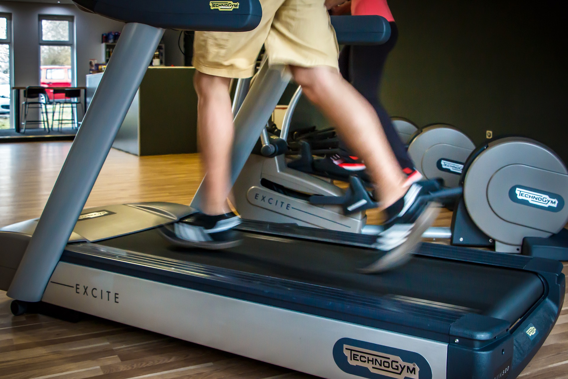
Is Pedaling Backwards on the Elliptical Machine Bad for Knees?

Pedaling backwards on an elliptical machine may be just what the doctor ordered for your knees; there’s no need to think that this might harm your knee joints in any way.
The research was presented at the American College of Sports Medicine’s 58th Annual Meeting and 2nd World Congress on Exercise in Medicine® in Denver in 2011.
In terms of cardiovascular fitness and muscle strength, study subjects who pedaled backwards on the elliptical (and walked backwards on a treadmill) did better than those who moved forward.
Here is what the lead author of the study, Elmarie Terblanche, PhD, states:
“Participants who used backward locomotion showed significantly greater gains in quadriceps and hamstring strength.
“Additionally, they had greater aerobic capacity than the forward-locomotion group.”
The subjects had a variety of knee injuries and were randomly assigned to backwards and forwards pedaling groups.
They completed 24 supervised sessions that included exercises for strength, flexibility and balance.
The backwards pedaling group averaged a 9 percent greater capacity in aerobic fitness.
“We say, do it backward!” says Terblanche, referring to rehabilitation routines for those with knee injuries.
The deduction that I can make as a personal trainer is that people with healthy knees can benefit from pedaling backwards on the elliptical machine as well.
This is not something that should be reserved for just those with “bad knees” or who are overweight or older.
Even young athletes should do this, as it provides a unique challenge.
Backwards pedaling feels less intimidating to most people, than does walking this way.
Nevertheless, next time you’re on the elliptical, go backwards for a few minutes here and there out of your session.
For greater quadriceps recruitment, sink down a bit (lower your center of gravity).
Important: While in the lowered position, try to keep back straight!
Maintain balance by lightly keeping fingertips slightly contoured to the rails; do not outright grab the rails.
Because if you do, this will subtract workload off the legs, which will defeat the purpose of this type of training.
If you decide not to be lowered, then don’t hold on at all.
This will force your core to work to keep balanced!
Lorra Garrick has been covering medical, fitness and cybersecurity topics for many years, having written thousands of articles for print magazines and websites, including as a ghostwriter. She’s also a former ACE-certified personal trainer.
.
Source: acsm.org/about-acsm/media-room/news-releases
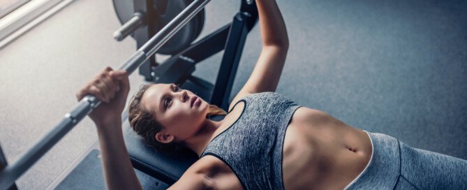
10 Bench Press Mistakes that Women Make

There’s a lot of info out there on the mistakes men make with bench pressing, but what about women? Yes, there is a difference.
More and more women are taking up the bench press, and this means more and more mistakes being committed.
So I will set things straight so that women can focus on doing the bench press right, and this includes not just technique, but information about this compound exercise that many women have wrong.
Common bench press mistakes women make (no particular order):
#1. Avoiding this effective exercise altogether out of fear it will bulk up their chest. Believe it or not, some women still think that bench pressing will give them a chest just like their husband’s.
Though women who compete in this exercise and lift literally 300, even over 400 pounds (Becca Swanson set a world record with a 551-pound lift in 2006) don’t exactly have the physiques that most women dream of, we’re not talking about training to break records.
This is about performing this multi-joint exercise as part of a fitness and toning regimen.
In order for a woman to start looking more like a man as a result of bench pressing, she’d have to be lifting as much as a man — and that’s men who are strong!
Men just starting out with this exercise may still look scrawny, even though they’re lifting 150 pounds!
You will not “bulk up” lifting the range typical for females: 45 pounds to barely 100.
At health clubs, it’s rare to see a woman benching what would be considered a lot of weight, and even then, they aren’t anywhere near bulked up.
Let’s crush this myth once and for all. Becca Swanson (5-10) weighs 240 pounds. She couldn’t weigh this much without a special dietary regimen to support it.
#2. Under-loading the weights out of fear of bulking up. See #1 above.
#3. Overloading the weights and then bringing the bar down only part way, but then believing they actually lifted all that weight.
Bringing the bar down only half way is not the same as all the way, and you’re fooling only yourself. This is a bad habit that can and should be broken.
#4. Thinking that this popular routine will make their breasts larger. If this were true, it would spread like wildfire and put breast-enhancement surgeons and manufacturers of “breast creams” out of business.
Why would a woman spend $5,000 on breast implants when she could just build up her breasts with the bench press? Weight-lifting does not build up fat — and breasts are made up of fatty tissue.
#5. Believing that this routine will shrink their breasts. Trust me, this won’t happen.
This concern was presented to me by several of my female clients when I was a personal trainer. “Will never happen,” I’d tell them.
#6. Not doing rotator-cuff exercises. Women need to realize that the bench press recruits the rotator cuff muscles and their tendons, especially as the weight becomes heavier.
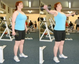
Dumbbell side lifts will warm up the rotator cuff. Source: GeorgeStepanek
Rotator-cuff-targeting exercises should be done to guard against injuring these tendons.
#7. Arching their back while straining to push up a weight. This defeats the purpose, because the back arch causes other muscles to get in on the act.
Bench pressing is for the chest/shoulders/triceps, not the back. Plus, the back arch can harm the back. Place your feet flat on the bench to discourage back-arching.
The exception is if you’re training for a powerlifting competition, in which an extreme back arch is very common because it reduces the distance the bar has to travel.
But again, if you’re bench pressing for fitness and strength, keep your back flat on the bench.
# 8. Not being aware that they’re pushing the bar up crooked. This is most prevalent when an individual uses just the Olympic bar (only 45 pounds), or an even a lighter bar.
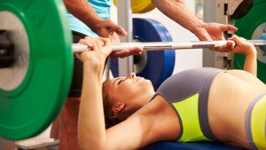
#9. Not asking for a spotter to help with heavier lifts. Many men will be more than happy to spot a lady with a bench press, even if she doesn’t look like Pam Anderson.
#10. Believing that the bench press is the only means to achieve results, be they muscle-toning, increased upper body strength or firmer arms. This exercise can be uncomfortable, even at lighter weights.
It is said that Arnold Schwarzenegger hardly ever, if at all, did this exercise on a flat bench, because of the injury risk to the rotator cuff. Instead, he used an inclined variation.
Women need to realize that the bench press is not the only means to an end; other exercises will do just as well, such as incline, dumbbell and horizontal chest presses.
Women should never assume they are missing something if the bench press is not part of their workout regimen.
Lorra Garrick is a former personal trainer certified through the American Council on Exercise. At Bally Total Fitness she trained women and men of all ages for fat loss, muscle building, fitness and improved health.
.


