

Traits of Women Who Are Abuse-Proof

It’s not always luck that some women never end up with an abusive partner; some really are abuse-proof.
We can’t put every woman into the same box and say there’s no such thing as an “abuse-proof” woman.
Have you ever wondered why some never end up with an abusive man? Is it luck? Is it because she never encountered him? For some, yes. But not for all women.
“Yes, some women are essentially abuse-proof,” begins Tina B. Tessina, PhD, a licensed psychotherapist in Southern California who counsels individuals and couples, and is author of over a dozen books including “It Ends With You: Grow Up and Out of Dysfunction.”
This doesn’t mean these feisty, self-assured women are capable of physically overpowering most men.
In fact, a 5’2, 105 pound woman may begin dating a man, and after a few weeks, they get into a disagreement about which restaurant to go to. He shoves her into a wall, then grabs her arm and flings her onto the bed.
Some women will continue dating this man and may even marry him, thinking his violent behavior was her fault, or that she could “change” him.
On the other hand, the abuse-proof woman, all 105 pounds of her, will get off the bed, shake herself off, and if it’s his house, she’ll immediately leave and have NO interest whatsoever in ever seeing him again. And she’ll stick to her guns about that.
Now, if the assault occurred at her house, she’ll figure out a way to get rid of him without risking her safety, then never have anything to do with him again.
That’s abuse-proof: Some women have a “one strike and you’re out!” policy.
What makes a woman abuse-proof?
“Women who grew up in an atmosphere of respect and caring are much more likely to be able to tell a bad guy from a good guy,” says Dr. Tessina, who counsels abused women.
Being abuse-proof is more likely when a girl grows up with a caring, loving father who respects her, her mother and other women in her life. This girl will grow up having expectations of how a man should treat her.
“Women who feel able to take care of themselves will also be more abuse-proof,” adds Dr. Tessina.
If a woman has earning power and family support, she is less likely to be drawn to an evil man who promises, “I will take good care of you.”
Not that such promises always mean trouble; plenty of wonderful men tell their girlfriends this, and then follow through for the next 50 years of marriage.
But Dr. Tessina also says, “If a women feels too dependent on a man to take care of her, she is vulnerable.”
Being abuse-proof begins with recognizing tell-tale signs of a controlling, wicked man. “Women who realize that even small incidents of abuse are unacceptable won’t stick around for more,” explains Dr. Tessina.
“When I talk to abused women in my counseling practice and teach them what to look for, they realize the signs were there from the beginning.”
It is never justified to criticize a woman who comments that she is abuse-proof.
Again, this is about the “one strike and you’re out!” policy.
Those who live by this policy aren’t the ones who make the news, so this creates the illusion that it’s an impossible feat for any woman anywhere to be immune to allowing a man to chip away at her self-esteem.
Women deserve more credit than to be lumped into the same cookie cutter box.
 Dr. Tessina appears frequently on radio, TV, video and podcasts, and has been in private practice for over 30 years.
 Lorra Garrick has been covering medical, fitness and cybersecurity topics for many years, having written thousands of articles for print magazines and websites, including as a ghostwriter. She’s also a former ACE-certified personal trainer.
.
Top image: ©Lorra Garrick

Do Children with Down Syndrome Suffer?

Almost every article I’ve ever read about Down syndrome has the word suffer.
These days, though, journalists and bloggers are more aware of the descriptors they use when referring to someone with Down syndrome.
But there definitely was a time, years ago, when you could bet that most articles that contained a reference to someone’s child or teen who had Down syndrome — used the words “suffers from.”
It goes as follows: John Doe, who suffers from Down syndrome…or, it could go like this: Jane Doe has a young daughter who suffers from Down syndrome.
Non-journalists do it too, in that they think a child or adult with Down syndrome “suffers.”
I’ve written several articles for print publications about a teen girl with Down syndrome — not a girl who suffers from Down syndrome. I wrote it out as: She has Down syndrome.
She had earned a black belt in karate as a teen, which was why I wrote about her.
She had gotten her driver’s license, and her parents had even bought her a car.
To say she suffers from anything doesn’t fit at all. She eventually got a part-time job at a clothing store. Kind of doesn’t quite meet the criteria for “suffers.”
Of course, there are people with Down syndrome who have major struggles including heart problems and deafness.
But I’ve found that in many of the referenced articles, the author didn’t know much about the child with Down syndrome — only that the adult they were talking about had a child with Down syndrome.
Often, the child’s name wasn’t even mentioned. The author had taken it upon themselves to say they “suffered from.”
Why not just say, “He has Down syndrome”?
I don’t live with them, nor do I see them often enough to know their lifestyle circumstances.
So where would I get off, then, using the word suffers in my articles about someone with Down syndrome?
What’s amazing is that the parents of kids or adults with Down syndrome let journalists get away with this.
Don’t parents ask to read an article first, before the writer sends the final version off to the editor?
You’d think that any parent reviewing an article, in which info about their child with Down syndrome is included, would be quite irritated at that word suffers.
A good journalist will replace “suffers” with a kinder word upon request.
The irony is that sometimes, the parents will talk about what an angel her child is, how loving this child is, how everyone’s her friend, how she’s always smiling — after the writer says this child suffers from Down syndrome.
If you believe someone thinks your child is suffering, ask them, “When’s the last time you saw someone with Down syndrome who appears to be suffering?
“In fact, don’t they always appear to be the happiest people in the room?”
Then be silent and gloat in the pride of making the skeptic realize his or her blunder.
Or, you can say, “Have you ever seen a disgruntled child with Down syndrome?”
Children with Down syndrome who are very high functioning know they are “different.” But even THEY seem to have a happy disposition as well.
“Look into my child’s face next time you see her, and tell me if you see any suffering,” you might say to the skeptic.
Parents say their kids with Down syndrome — children as well as adults — have a sunny, content disposition, enjoy affection and have brought tremendous joy to the family. Many have full-time jobs. Who’s suffering?
When I hear the word suffer, I think of someone who lives in pain, experiences mental anguish, battles drug addition, has debilitating lupus, diabetes or some other medical sickness, lives with an alcoholic, etc.
People suffer from paralysis, cancer, depression and schizophrenia.
Though nobody wants their newborn baby to be diagnosed with any genetic anomaly, at the same time, I doubt anyone with this condition would describe themselves as suffering from it.
This is why many people with the condition say they have Up syndrome.
Lorra Garrick has been covering medical, fitness and cybersecurity topics for many years, having written thousands of articles for print magazines and websites, including as a ghostwriter. She’s also a former ACE-certified personal trainer.
.
Top image: Freepik.com

Should You Stay with Your Dog when It’s Time to Put it Down or Leave?

When it’s time to put your dog down, should you stay with the dog during the process, or leave the room before the dog starts drifting off towards doggie heaven?
I have given this some thought, regarding my grand White German Shepherd, and I’ve made a decision:
I’m staying right in the room with him, and I will be the last thing he sees before doggie heaven calls him home.
There’s an easy way to make this decision. You do it by fast-forwarding to that most inevitable future, where your beloved dog is ready to be put down.
It will be one of the most dreadful days of your life, and you certainly don’t want to make any regretful decisions. So, imagine that awful day has arrived to put down your dog. There are one of two outcomes:
#1. Stay with the dog as he is being put to sleep.
This will be very trying, as you watch the dog’s eyes, which are watching you, slowly start closing.
This happens after you witness the vet inject your pet and do whatever else is necessary.
You are literally watching your dog dying, and not just dying, but being put to death. To say a pet is put down or put to sleep softens the blow to the master.
When the ordeal is over, you walk out of the room, trembling, because you just witnessed your best friend’s last breath. You saw your dog die.
Forever in your mind will be that image. The last memory you have of your dog is of it dying, and then being dead. Can you live with that?
#2. Say your last goodbye to the dog while it’s still fully conscious.
The dog is watching you as you slowly back out of the room. You wonder if the dog knows it’ll never see you again. You hurry outside to the parking lot and burst into tears.
The last memory of your pet will be when it was alive and watching you. Can you live with that?
Which option is more regrettable? Let’s dig deeper. Is this about you, or the dog?
It should be about the dog. What would your dog want? Pretend you yourself are dying.
What would YOU want? You’d want your loved-ones at your side, holding your hand, seeing you through to your last breath.
This is what your dog would want. If you choose #1, the last memory of your dog will be as it’s being put to sleep.
However, you will have comfort knowing that right up to the last minute, you were at the dog’s side, hand on its head or hand holding its paw.
You were there. Your pet felt your protection right up to the end. You helped your best friend make that transition to doggie heaven. You’ll never regret that.
If you choose #2, you may initially think this was the best choice, because it won’t leave any memories of a dying animal, just an awake, alert dog.
But give it a few days; something awful will begin sinking its claws into you:
The realization that you were not there.
From your dog’s point of view, you abandoned him.
You left him alone with those people who put him to sleep. You didn’t stay at his side. You chickened out.
You’ll regret that for all time.
Lorra Garrick has been covering medical, fitness and cybersecurity topics for many years, having written thousands of articles for print magazines and websites, including as a ghostwriter. She’s also a former ACE-certified personal trainer.
.
Top image: Shutterstock/Sergey Nivens

How to Get Your Dog to Stop Passing so much Stinky Gas

Phewwww, does your dog’s passing gas stink up an entire room? Here is how to get that undern control — by a veterinarian.
Why does your dog, though, pass gas so often in the first place?
“Flatulence comes from an excess of gases that are in the intestinal tract,” says Michel Selmer, DVM.
“These may represent air that has been swallowed; produced in the biochemical process of digestion; or diffusion from the bloodstream, or gases produced by bacteria that populate the intestinal tract.”
Dr. Selmer, who’s with The Caring Vet in NY, explains, “Over 99 percent of the gases that pass from the intestinal tract are odorless; gases with objectionable odors are typically those containing hydrogen sulfide.”
Passing gas does not mean a problem with health.
“A surprising amount of air is swallowed with the simple act of eating,” says Dr. Selmer, “and if this is not burped out, it must exit through the other end.”
Additionally, “The amount of air swallowed tends to be increased when dogs feel they must eat quickly.”
The brachycephalic breeds (such as an English bulldog or Pug) tend to use their mouth more to breathe, rather then the nose, which is short. Air that’s swallowed usually doesn’t have a bad odor.
But what about really stinky gas passing?

Shutterstock/Anatoliy Karlyuk
“The really stinky gases are produced by large intestine bacteria,” says Dr. Selmer.
“Dietary fiber in pet food is not easily digestible by the pet’s own enzyme systems; but is digested by the gas-producing bacteria of the colon.”
These fibers get broken down, producing gas. “A diet heavy in fibers tends to favor these gas-producing organisms,” continues Dr. Selmer.
“The more supportive the intestinal environment, the more bacteria there will be; ultimately more gas will be produced.”
Solution to Passing Gas Less Frequently in Your Dog
No. 1: Lower the fiber amount in the dog food. Read ingredients for anything fibrous. Check the nutrition label if a fiber content/serving is listed. Cut back on grains.
No. 2: It’ll help of your dog can eat more slowly.
Only one meal a day will cause rushed eating.
Try multiple servings but with smaller amounts; this can cut down on passing gas.
 Dr. Selmer offers conventional Western plus holistic veterinary medicine, traditional Chinese veterinary medicine including acupuncture and herbal therapy, and integrative medical care for dogs and cats.
Lorra Garrick has been covering medical, fitness and cybersecurity topics for many years, having written thousands of articles for print magazines and websites, including as a ghostwriter. She’s also a former ACE-certified personal trainer.
.
Top image: Shutterstock/STUDIO GRAND WEB

Dogs with Sensitive Stomach Must Avoid THESE 10 Foods

From a veterinarian’s mouth are the 10 foods dogs with sensitive stomachs need to avoid eating.
“Sensitive stomachs in dogs can result from food intolerance,” says Dr. Michel Selmer, DVM, with The Caring Vet in NY.
10 Foods Never to Give a Dog with a Sensitive Stomach
According to Dr. Selmer, here are at least 10 food items (no particular order) that you should never give a dog with a sensitive stomach.
High fat foods. These foods are “difficult to digest and can cause inflammation of the pancreas,” notes Dr. Selmer. There are two exceptions: ground beef (low fat) and peanut butter.
Chocolate. Dogs cannot metabolize the theobromine in chocolate. Chocolate can cause seizures in dogs.
Coffee and tea. These are as bad as chocolate.
Raisins and grapes. These can lead to kidney failure.
Nutmeg. Seizures can result.
Raw eggs. Salmonella may be a problem.
Onions. Dr. Selmer says onions “are not advised, as they can interfere with blood circulation.”
Macadamia nuts. Dr. Selmer says these “can cause dogs to have tremors and lead to paralysis.”
He continues, “Other food cautions include moldy foods, yeast dough and fruit pits. Many pits contain cyanide.”
For stomach-sensitive dogs:
“Food intolerances can result from dyes, preservatives, contaminants or even natural proteins in the food,” explains Dr. Selmer.
“The solution is the feeding of a ‘pure’ diet, ideally home-cooked food made with carbohydrates and proteins that are new to the patient.
“Recently a new approach has been introduced that uses therapeutic diets made from hydrolyzed proteins. This means a conventional protein source is used; the protein is broken down into molecules too small to excite the immune system.”
Home-cooked Foods for Dogs with Sensitive Stomachs

Rice. “Rice is a good carbohydrate for dogs,” says Dr. Selmer. “Brown rice is preferable, as it has more nutrients.”
Brewers yeast
Flour (e.g. soy flour)
Cod liver oil or flaxseed. Dr. Selmer says that in small amounts these add omega 3 and helps keep their coats shiny.
Garlic; “can help get rid of fleas and tapeworms as well as fight infection; caution; large amounts can cause illness in dogs.”
Meats. Liver, beef, tuna, chicken, lamb. “It’s easier to add to food if the meat is ground.”
Dogs love peanut butter (who knew!), “and biscuits can be made with them by adding flour, bone meal and/or powdered milk, brewers yeast and carrots. Peanuts are one of the few nuts safe for your dog.”
He endorses also broccoli and spinach, “but they need to first be put through a food processor to aid in digestion.” (Don’t give broccoli in large amounts.) Add bone meal for calcium. “Raw meaty bones are a good source of calcium. “Powdered milk is also a popular ingredient in dog food.”
You now know how to manage the food intake of a dog with a sensitive stomach.
Dr. Selmer offers conventional Western plus holistic veterinary medicine, traditional Chinese veterinary medicine including acupuncture and herbal therapy, and integrative medical care for dogs and cats.
Lorra Garrick has been covering medical, fitness and cybersecurity topics for many years, having written thousands of articles for print magazines and websites, including as a ghostwriter. She’s also a former ACE-certified personal trainer.
.
Top image: ©Lorra Garrick

Don’t Let Dog’s Strep Throat Symptoms Fool You

Can YOU OR your vet (!) properly recognize strep throat symptoms in your dog?
My parents’ dog almost died from a strep throat, thanks to a misdiagnosis by a veterinary neurologist who was treating him for a brain tumor.
The ER vet at a prestigious veterinary clinic where the neurologist was based examined the German shepherd and couldn’t come up with anything definitive; the animal stayed overnight.
Next morning and $1,300 later, we were informed by the vet neuro that the deterioration was due to the brain tumor’s fluid buildup and/or the tumor itself.
He was treated with a diuretic and anti-inflammatory via IV feed.
The GS perked up somewhat for the next eight hours, then relapsed; for the next five days he outright deteriorated: increasing appetite loss; increasing listlessness and weakness; excessive sleep; depressed; glazed look in eyes.
Our pet was eventually sleeping all the time except when the urge to urinate was so overwhelming that he forced himself up and stood at the door; he was also drinking huge amounts of water.
By then, he had had one visit with a new veterinarian who uses alternative treatments for cancer.
Over the phone I described the dog’s symptoms to the doctor, and he promptly diagnosed strep throat!
“Bring him in tomorrow morning at 7:30,” he said. Next morning the German shepherd’s fever was over 104 degrees.
He stayed a few days and was “rebuilt,” explained the doctor, who told us he sees strep throat in dogs all the time.
General Symptoms of Strep Throat in a Dog
Lethargy; a depressed demeanor; physical weakness; glazed, glossy eyes; appetite loss/slower eating; increased water intake; fever; unwillingness to get up at commands; extremely excessive sleep.

In advanced stages of strep throat, a dog will be virtually unresponsive to commands; will need help getting on all fours; may even need to be carried to get from point A to point B; refusal to eat.
Early Symptoms of Strep Throat in Dogs
Subdued nature; glazed or hazy eyes; lack of gusto when eating; picky eating; increased water intake; listless gait; hacking or gagging sounds; upchucking or spitting up; diminished interaction with owners.
The German shepherd began showing early symptoms of strep throat a second time, after fully recovering from his first strep experience.
Because the strep this time was in its early stages, the dog was given a penicillin injection. His full appetite returned that day.
Next day he had a second injection; we took home penicillin in tablet form plus two more drugs to fight the infection, and he was eating like a vulture.
Our vet said, “I see 10 dogs a day with strep.”
There are mulitple strains of the Streptococci bacterium. “Many species of Streptococci are normal residents of the skin, urogenital tract, nose and mouth,” points out Dr. Michel Selmer, DVM, with The Caring Vet in NY.
“People are the natural reservoir hosts for Group A Streptococci,” continues Dr. Selmer. “Group A Streptococci causes ‘strep throat’ in people. Dogs and cats are not reservoirs for Group A.”
Dr. Selmer adds, Group B Streptococci have occasionally been associated with neonatal infections, endocarditis, endometritis and other mixed infections in dogs. Group G Streptococci are normal residents of the skin and mucosa of dogs, responsible for most infections in dogs.”
Canines infected with Group G “May have a history of recent trauma or bite wounds, or may have nonspecific signs (lameness, respiratory signs, urinary signs),” continues Dr. Selmer.
“Most dogs develop a rapidly progressive, severely painful cellulitis. The pet’s limbs are most often affected.
“The dogs become depressed and shocky. Fever, depression, hypotension low blood pressure] and shock may also develop in infected dogs, in the absence of fasciitis.
“Acute pulmonary infections with Streptococci appear to be the main cause of the streptococcal toxic shock syndrome.
“Most dogs with toxic shock syndrome (with or without fasciitis) die or they are euthanized due to the severity of their signs.”
How can strep throat be prevented?
Dr. Selmer says, “Because most Streptococci are normal residents of the skin and mucosa, animals are universally exposed to these bacteria.” A compromised immune system is a risk factor.
“Routine prevention involves good sanitation and hygienic care of wounds and other non-infected lesions.”
Dr. Selmer offers conventional Western plus holistic veterinary medicine, traditional Chinese veterinary medicine including acupuncture and herbal therapy, and integrative medical care for dogs and cats.
Lorra Garrick has been covering medical, fitness and cybersecurity topics for many years, having written thousands of articles for print magazines and websites, including as a ghostwriter. She’s also a former ACE-certified personal trainer.
.
Top image: ©Lorra Garrick

Reasons a Domestic Dog Howls When It’s Never Done So Before

If your dog has always howled, you may not give it much thought as to why; after all, domestic canines are descendants from wolves.
But what if your dog suddenly starts howling out of the blue, for no apparent reason?
“Usually dogs howl in response to a sound stimulus. In the wolf population it’s a calling sound or a response to a calling,” says Mary Remer, certified pet dog trainer, breeder and licensed AKC judge, having trained more than 20,000 dogs in her 30-year career.
If you watch Animal Planet enough, you’ll sooner or later see a show about a person who spends a lot of time with wolves.
This person will be perched on a rock, for instance, and will start howling.
Before long, the wolves that he or she has befriended will gather around this individual and start howling as well. It’s a social event. Howling means, “Come join me.”
Remer continues, “In domestic dogs, we tend to see howling more in response to a sound, such as a fire alarm, or sometimes musicians report that their dogs howl when the instrument they play is at a certain note or pitch.

“Sometimes dogs howl if there is a particular song on the radio and may react to a pitch or a note and the dog responds with a howl.”
My parents’ German shepherd would let out a sustained, forceful howl whenever the doorbell rang, and then it was followed by a series of guard-dog-like barks.
Remer adds, “Some dogs howl because they’re lonely.”
In my parents’ neighborhood is a husky that’s often left out in the yard for many hours on end all by itself; this poor husky has howled incessantly and sounded like one miserable animal.
The howling was nonstop and it was a wonder this stately husky didn’t damage its vocal cords.
My mother finally put several complaints into the homeowners’ association and the howling has since ceased.
No pet should be left outside alone for literally eight hours, like this lonely husky was.
“Some dogs howl as part of locating their prey,” says Remer. “Certain hound breeds will howl as an indication of locating their prey. Some hounds such as treeing hounds and coon hounds howl to let the human hunter know its location.
“Bassets, beagles and fox hounds have a howl-like noise known as a cry when they are hunting and will also make the sound in an aroused state anticipating hunting.”
And sometimes, it’s just not clear why a dog will start howling out of the blue. My parents’ German shepherd, as mentioned, normally let out a single howl whenever the doorbell rang, followed by aggressive barking.
But not long after being diagnosed with a brain tumor, the German shepherd began howling while in the car, even though there were no apparent auditory stimuli around, or people around, while the car was parked in the lot; or while I was driving the car.
The German shepherd had been in cars since puppy-hood, so it was a mystery why one day he began emitting a more passive, and very prolonged, type of howl; it was a very charming howl that I enjoyed listening to, yet at the same time, it made me wonder if the disease was affecting his thinking, even though he seemed to be improving from treatment.
This magnificent German shepherd not long after made his journey to the Bridge.
In conclusion, howling is definitely a communication device among domestic dogs.
 Mary Remer is founder of What a Good Dog, a dog training and behavioral facility.
Lorra Garrick has been covering medical, fitness and cybersecurity topics for many years, having written thousands of articles for print magazines and websites, including as a ghostwriter. She’s also a former ACE-certified personal trainer.
How to Convince Someone It’s Time to Euthanize Dog with Cancer
It’s alarming to have a healthy dog put down just because it lost one leg in an accident, but cancer is a whole different ball game.
Should a dog with terminal cancer be put down?
My mother recently had her dog, who had cancer, put down. Long before her pet became sick, my mother, from time to time, had commented, “I can never put a dog down.”
These comments were prompted by things she might have seen on TV, or hearing about a friend who had a dog put to sleep.
Never in her craziest dreams did she ever imagine that she’d have to make this decision about her own German shepherd.
The dog had brain cancer and the day of diagnosis, my father said the animal should be put down.
At this early stage of the game, I was completely against this idea; we should try treatments first to see how much quality life we can get the German shepherd. My mother agreed.
However, I also knew that there might come a time that the cancer would start winning the fight, and that putting the dog down would be the best decision. It would take a mountain to convince my mother of this.
So early on in the dog’s cancer treatment, I began priming my mother for this decision making process.
Convincing someone to put a dog with cancer to sleep begins with bringing up the topic early on in the treatment.
#1: Early on in cancer treatment, begin talking about euthanasia — not in terms of that it should be done, but in terms of that one day, you may have to consider it.
#2: Point out that two conditions must be met before deciding to put a dog with cancer down:
A) All treatment options have been tried, given time to work, and exhausted, and B) The animal is no longer improving, not even stabilized, but is clearly deteriorating. Emphasize that these two conditions must be met first.
#3: From time to time, bring up the topic of euthanasia. You may feel there’s never a right time.
I did this periodically with my mother, even though each time, she was resistant, though I could tell that a tiny part of her deep inside, agreed with the idea of those two conditions being met.
I was staying with my parents while their dog was getting treated for the cancer, because my parents were not able to administer the 12 medicinal injections a day (six in the morning; six at night).
Furthermore, the menagerie of pills to give the GS was overwhelming to her.
Furthermore, I wanted complete control of the dog’s diet. Though my parents owned the German shepherd, he was very dear to me.
#4: Point out that when the dog starts going downhill, a point of no return, it’s time to put him down.
Otherwise, here is the alternative: Waking up to see a dead dog. I said, “Do you really want to come downstairs and be met by a stiff dog, he’s lying on his back, all fours sticking straight up, tongue hanging out, and you smell the beginnings of decomposition?”
Add: “Do you want your last memory of him to be that of death? Furthermore, if you don’t have him put down after those two conditions are met,
Dad and I will get charged with the task of picking up a dead dog and loading him into the car trunk.
I can’t go through that a second time.” (My parents’ first dog died of old age and my father and I performed that ugly task.)
Add: “Just think what would happen if you allow nature to run its course: Every night when you go to bed, you will be in a state of anxiety, wondering if next morning will be THE morning. You will not be able to sleep.
“You will toss and turn. Then come morning, your heart will be pounding as you make your way downstairs, wondering what you will see.”
Add: “If you put the dog down before the cancer takes him, you will spare yourself of a far worse possibility:
The animal starts convulsing in the middle of the night, spitting up its guts, thrashing around, then dies. It’s 3 a.m. What are you going to do?”
When my parents’ dog began deteriorating, I picked up on it before they did.
But eventually, my mother was talked into agreeing to have the dog put to sleep.
It broke her heart, but I convinced her that the alternative (the pet convulsing in middle of night, howling in pain, dying on the kitchen floor), was far uglier.
Lorra Garrick has been covering medical, fitness and cybersecurity topics for many years, having written thousands of articles for print magazines and websites, including as a ghostwriter. She’s also a former ACE-certified personal trainer.
.
Top image: Shutterstock/Sergey Nivens

German Shepherd Ear Infections Near Brain: Beware of Wrong Diagnosis
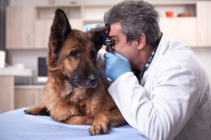
My parents had German Shepherd that has always seemed prone to ear infections.
I was house sitting the German Shepherd while my parents were away. Another ear infection was flaring up.
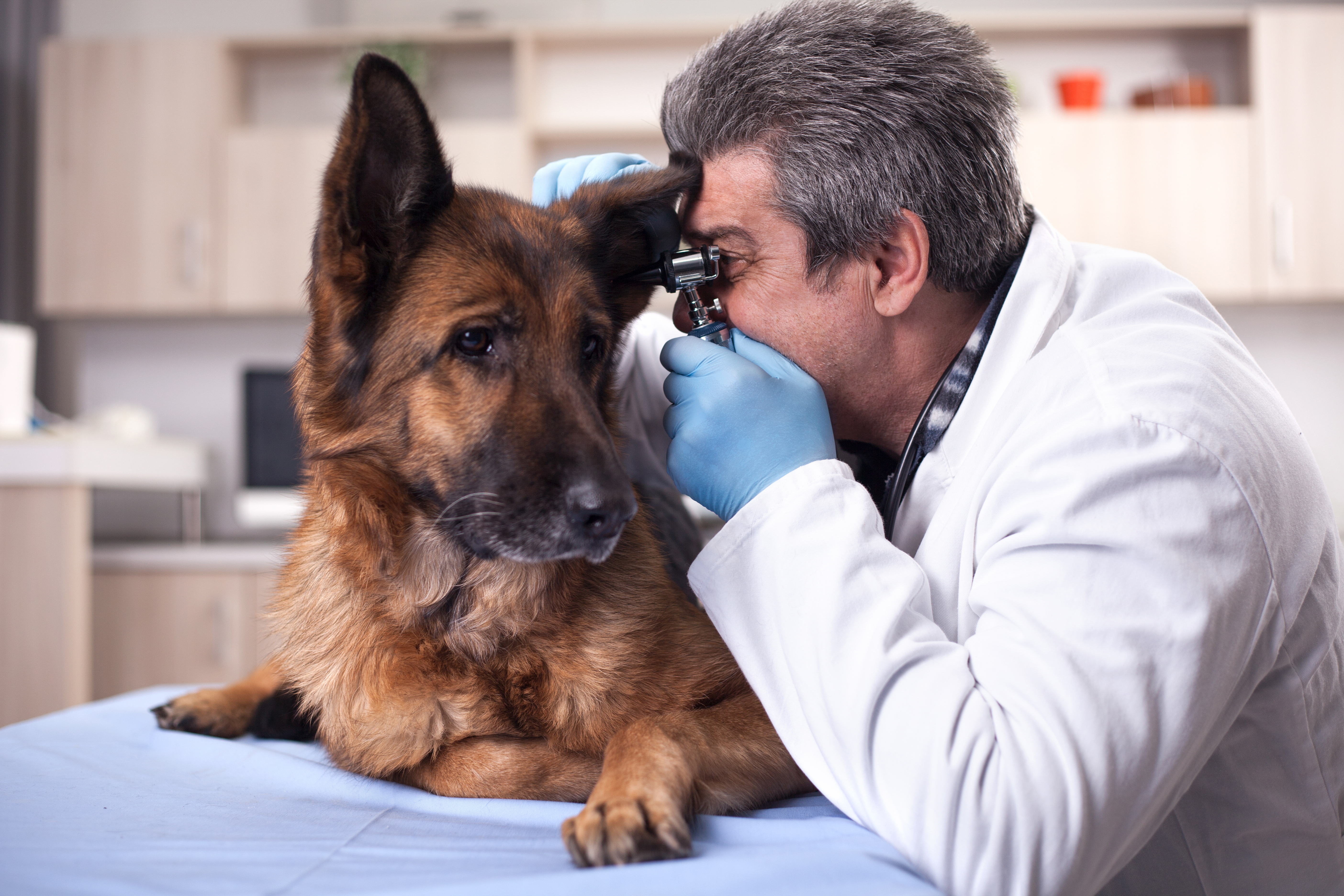
The ear infection was causing the dog to tilt his head and slowly shake it. I took the dog to the emergency vet on duty Sunday evening, who diagnosed a middle ear canal infection. This vet didn’t seem overly concerned about the dog.
I had witnessed a few vet techs cleaning out the ear; one vet tech held the muzzled dog in a tight embrace so that he could not get away. The other vet tech was wiping inside the ear with some little towelettes.
Meanwhile, the German Shepherd cried out in pain. The young vet, who had lackluster bedside manner, gave me a bottle of soap, antibiotics and pills.
When my parents returned, the ear infection seemed to be clearing up, in that the head tilt had disappeared by the day after I had taken the German Shepherd in.
But the dog was still scratching, though less, and the inside of his ear looked horrible, as always.
It was crusty and brown, not fleshy and pink like the other. Furthermore, getting the eardrops into his ear was an almost impossible task.
I had to muzzle the German Shepherd, and he kept walking away from me when he saw me raise the bottles.
He’d snarl like I’d never heard him before. He even tried to take a few nips.
My brother is a chemist and was shocked that one of the bottles was a soap that contained acetic acid. He said this was probably causing the dog searing pain.
My parents decided to try a completely new veterinary hospital, a vet specializing in dog ear infections.
They dropped the German Shepherd off there and an hour later, received a phone call from the vet.
The dog had a raging ear infection deep in the ear canal. His eardrum was ruptured.
- The infection was about one-quarter-inch away from his brain.
- There was severe inflammation.
The quack vet at the other clinic never said any such thing. This new vet said the German Shepherd had to be anesthetized so that he could get deep inside his ear and clean everything out.
Next time I visited my parents, there were half a dozen prescription bottles on the counter.
The dog had to take 15 pills a DAY, plus get eardrops daily — they took the dog every day to the clinic so that the vet could administer the drops.
About a week later, the ear looked practically brand new; nice and pink and fleshy, whereas, prior to that, it was chronically brown and crusty.
We believe that the German Shepherd never had recurring ear infections.
Rather, he had just one ear infection, and the first veterinary hospital failed to take care of the problem.
They just merely treated the symptoms, so that the infection was never eradicated.
There was always bacteria there, and once the feeble treatment stopped, the bacterial colony would have an opportunity to grow huge again, causing the dog to scratch relentlessly at his infected ear, and hiss in pain.
Trust your instincts. My mother had visited the first vet shortly after I had done so, and was totally put off by the vet’s blasé, indifferent demeanor. The vet had been the same with me as well.
If your dog has a “recurring” ear infection, consider that it’s been the same infection all along, and that the current treating vets are doing a quack job.
 Lorra Garrick has been covering medical, fitness and cybersecurity topics for many years, having written thousands of articles for print magazines and websites, including as a ghostwriter. She’s also a former ACE-certified personal trainer.
Top image: Shutterstock/VP Photo Studio








Sick Dog Starting to Die? Wait; May Be Treatable Strep Throat
Don’t put down your “dying dog” yet; may be treatable strep throat.
Is your sick dog’s dying process starting and you’re making arrangements to put the dog down? Does death seem imminent for your cancer-stricken pet?
Don’t call the vet or Humane Society yet to have the dog put down; a strep throat infection can mimic dying, in that it can seem like death will occur within a day or even hours – death from cancer or whatever your dog was originally diagnosed with.
Don’t be too hasty with plans to put your dog down until you get it checked for strep throat. This happened to my parents’ dog.
The animal was diagnosed with a brain tumor about three weeks prior to when he took a sudden turn for the worse. He was taken to the ER of the diagnosing facility.
The dog had suddenly become listless and lethargic. The treating neurologist said it was fluid buildup in the brain from the tumor.
The German shepherd was kept overnight, and $1,300 later, was sent home all perked up, having been treated with mannitol, an anti-inflammatory, and furosamide, a diuretic, via IV catheter.
Come evening, the animal had regressed and had diminished appetite. Next day he was more lethargic, slept excessively and had diminished appetite. We thought it was side effects from chemo.
Next day he was worse, doing nothing but sleeping, not responding to familiar verbual cues, and exhibiting severe hind leg weakness.
He was growing increasingly out of it and appeared to be entering his “final” stage; it appeared that death was around the corner; that “this is it.”
We braced ourselves as the evening wore on. The dog refused to eat; his breathing was very rapid while he deeply slept; he had profound body weakness; his eyes, when open, lacked life.
“He’ll probably expire overnight,” said my father, who had suggested calling the Humane Society for someone to “come over with a needle,” after the dog collapsed in the garage (after we let him out the side door to relieve himself) and refused to budge.
Once back inside the house, he resumed sleeping. This is it, we all thought. By then, we had begun taking the animal to a holistic doctor.
We thought the new treatment wasn’t working; the dog’s ears wouldn’t even twitch when we announced familiar verbal cues that historically excited him.
We called the new doctor that evening; he said bring the dog to his clinic 7:30 am next morning; he suspected a strep throat.
Next morning: Diagnosis: Strep Throat. Here we were, thinking the dog was dying, had reached the end of his rope.
Many other people in the same boat would have taken their dog to the nearest shelter or vet to be put down.
Had we had the dog put down, we would have had a dog put down for a strep throat!
A strep throat is very treatable, though success is not guaranteed. But you don’t put a dog down for a strep throat!
Symptoms of strep throat in a dog can mimic the dying process: rapid breathing, excessive sleeping, not easily aroused, no interest in food or social interaction, glazed, half open eyes when not asleep, significant weakness, collapsing on hind legs, unsteady gait.
The German shepherd’s temperature turned out to be 104.7; normal is 101-102. The doctor diagnosed him within one minute of examining him.
Later in the afternoon, the doctor called to say that the animal was doing very well, and had been walked twice around the building, which includes a hill!
If your dog, that’s been diagnosed with a serious disease, seems to be dying or on its way out, consider having the animal examined for a strep throat before you arrange to have it put down.