Medical plans don’t include the mention of “routine” MRI screenings for a brain aneurysm.
And this seems pretty odd, given that often, the first symptom of such an aneurysm is a thunderclap headache soon followed by death.
Though you won’t find a recommendation for annual brain aneurysm screenings in your medical plan’s booklet, annual MRI’s are recommended for those individuals who are at high risk.
“The prevalence of brain aneurysm by radiographic and autopsy series ranges from 0.4 to 6.0 percent,” says Farhan Siddiq, MD, a neurosurgeon with University of Missouri Health Care.
Startling Statistic
“In the adult population, without any risk factors, two percent may have an unruptured aneurysm,” says Dr. Siddiq.
“The most effective and safe test for screening is an MR-angiogram.”
This MRI technique, known as magnetic resonance angiography (MRA), involves injecting a contrast dye into the patient.
The dye enhances the imaging, making blood vessels and any aneurysms within them appear clearly on the screen.
By highlighting these structures, MRA effectively reveals bulges or abnormalities in blood vessels, aiding in the diagnosis and assessment of aneurysms.
As simple as this seems, there’s a reason why this procedure is not part of one’s routine wellness checks as is, for instance, the mammogram, colonoscopy and cholesterol checks.
“Because of the high cost of MRA, along with the low frequency of aneurysms in the general population, routine screening would not be cost-effective,” says Dr. Siddiq.
What exactly is an aneurysm?
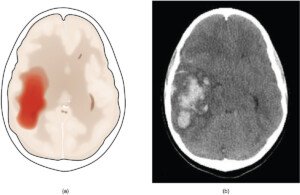
An aneurysm is a bulge or swelling in the wall of a blood vessel caused by weakness or damage.
This can occur in arteries throughout the body, not just the brain.
Aneurysms are often asymptomatic and may be discovered incidentally during imaging tests, such as a head scan, conducted for unrelated medical reasons.
While some aneurysms may remain stable, others can pose serious risks if they rupture, leading to internal bleeding or other complications.
Symptoms of an aneurysm in the brain include a drooping eyelid (ptosis), double vision and pupils of marked unequal size.

Ptosis
Dr. Siddiq adds, “Possibly, in the future, we may have better, faster and cheaper tests, which would make it more feasible for routine screening.”
An MRI is generally more effective at detecting brain aneurysms compared to a CT scan.
MRI provides detailed imaging of brain structures and blood vessels, allowing for clearer visualization of aneurysms.
This superior, very sensitive detail helps in diagnosing and assessing aneurysms more accurately.
High blood pressure and smoking are risk factors for a ruptured aneurysm.

Dr. Siddiq is fellowship-trained in endovascular surgical neuroradiology and vascular neurology from the University of Minnesota Medical Center. His areas of special focus also include brain aneurysms and carotid disease.
 Lorra Garrick has been covering medical, fitness and cybersecurity topics for many years, having written thousands of articles for print magazines and websites, including as a ghostwriter. She’s also a former ACE-certified personal trainer.
.


