

How Much Worse Is a Total Cholesterol of 1,000 than 500?

Can total cholesterol be SO high, 1,000 vs. 500, that anything higher would not make a difference in how at risk the patient is for a heart attack?
What about treatment options; would they differ? (more…)
Chest Pain from Anemia vs. Heart Attack: Symptom Comparison
What features of chest pain from a heart attack would not be felt from anemia?
A person may also have symptoms days before the actual heart attack. (more…)

Can Heart Palpitations Be Caused by Low Iron Levels?

Heart palpitations—the perception that the heart is skipping beats, making extra beats or beating erratically, has multiple causes.
Low iron levels can be associated with this situation, which ranges from annoying to frightening.
Suddenly feeling your heart doing flip-flops or a perceived “skipping” or “extra” beat can stop a person in their tacks and make them worry they’re about to have a heart attack.
Iron Levels
“Low blood iron levels, in themselves, do not cause palpitations,” says Morton Tavel, MD, Clinical Professor Emeritus of Medicine, Indiana University School of Medicine, and author of “HEALTH TIPS, MYTHS, AND TRICKS: A Physician’s Advice.”
“However, if low iron levels are associated with anemia, as they often are, the resulting fewer red blood cell levels carry less oxygen.
“Therefore, in attempting to sustain adequate oxygenation of all the vital organs, this may result in a rapid heart rate and the sensation of palpitations when one exerts him/herself, even at relatively low levels.”
Definition of Anemia
Anemia is when there are not enough red blood cells in a person’s circulation to deliver optimal oxygen to the body’s cells.
When red blood cell levels are too low, or when they do not work properly, the body’s cells receive less oxygen than they need.
This reduced oxygen delivery can interfere with normal cellular activity.
Common Symptoms of Anemia
Fatigue and weakness are common symptoms.
In addition to an irregular heartbeat, other symptoms of anemia include pale skin, a feeling of being lightheaded, dizziness, chest pain, headache and cold hands/feet.
Why do low iron levels result in fewer red blood cells?
The bone marrow needs iron, a naturally occurring metal, to produce hemoglobin.
Hemoglobin carries oxygen and is found in red blood cells.
Not enough hemoglobin means symptoms of anemia.
A blood test will determine if you have a low red blood cell count.
If so, your doctor will then seek out to find the cause and treatment.
In the meantime, your heart palpitations may also be caused by anxiety or even excessive caffeine intake.

Dr. Tavel’s medical research includes over 125 publications, editorials and book reviews in peer-reviewed national medical journals. He was formerly director of the cardiac rehabilitation program at St. Vincent Hospital in Indiana. mortontavel.com
 Lorra Garrick has been covering medical, fitness and cybersecurity topics for many years, having written thousands of articles for print magazines and websites, including as a ghostwriter. She’s also a former ACE-certified personal trainer.
.
Top image: ©Lorra Garrick

Can Tight Shoes Cause Permanent Peripheral Neuropathy?

Tight shoes can feel as though they are very gradually causing peripheral neuropathy if you wear them often.
In order for tight shoes to cause permanent peripheral neuropathy, you’d have to wear them way more than what you normally do. (more…)
What Does Sugar in the Urine of Non-Diabetics Mean?

Sometimes, sugar can find its way in the urine of people who don’t have diabetes.
This can make them wonder if diabetes awaits them around the corner.
What Sugar in the Urine of Someone Without Diabetes Might Mean
“Most likely it means nothing, but it could mean a problem in kidney function such that the kidney is unable to properly reabsorb glucose (in certain chronic kidney disease or a relatively rare genetic defect),” says Susan L. Besser, MD, with Mercy Medical Center, Baltimore; Diplomate, American Board of Obesity Medicine and board certified by the American Board of Family Medicine.
Glucosuria is the general name for sugar (glucose) in the urine in people who do not have diabetes.
In someone with healthy kidneys, glucose ends up in the urine when blood sugar gets abnormally elevated.
A rare condition called renal glycosuria results from an impairment of renal (kidney) function. If you’re non-diabetic but have excess sugar in your urine, you could have renal glycosuria.
Likely you’ll have no symptoms or ill effects. This is a benign condition in most cases.
In other cases there may be excessive urination and thirst.
The condition is inherited (genetic) and is not caused by lifestyle habits.
Dr. Besser also says, “Lastly, some medications may cause glucose to be spilled in the urine (although these medications are usually used to treat diabetes).”
These medications include: Canagliflozin, Dapagliflozin, Thiazide diuretics (e.g., hydrochlorothiazide), Corticosteroids (e.g., prednisone), Cisplatin, Ifosfamide, Tenofovir
Signs of Diabetes
- Unintentional weight loss
- Unexplained fatigue
- Unexplained hunger
- Excessive thirst and urination
- Problems with vision
About 90 percent of diabetics have the type 2 version, and though obesity is a risk factor, thin people can get type 2 diabetes.
This is because type 2 diabetes can be linked to genetic factors, insulin resistance or fat distribution—especially excess visceral fat around organs.
One need not have a conventionally fat body to have excess visceral fat. Chronic stress, poor diet, inactivity and certain medical conditions can also impair insulin function.
If your urinalysis shows that you have excess sugar in your urine, and you do not have diabetes, then ask your doctor what this may mean for your particular situation and medical history, and if any additional testing is called for.
 Dr. Besser provides comprehensive family care, treating common and acute primary conditions like diabetes and hypertension. Her ongoing approach allows her the opportunity to provide accurate and critical diagnoses of more complex conditions and disorders.
Lorra Garrick is a former personal trainer certified by the American Council on Exercise. At Bally Total Fitness she trained clients of all ages for fat loss, muscle building, fitness and improved health.
.
Top image: Shutterstock/Guschenkova

How Much Can Light Smoking Raise DVT Risk?

Could a smoking habit be light enough to no longer be a risk factor for DVT?
Or is the risk of deep vein thrombosis the same for light smokers as it is for moderate and even heavy? (more…)

Does Upper Arm Size Affect Blood Pressure Reading?

If you think that your big arm is affecting your blood pressure readings, you’re correct.
The size of your upper arm affects the blood pressure reading. (more…)

Can a DVT Come Back in Its Original Location?

If you’ve been treated for a deep vein thrombosis, is it possible that it could come back in its original location?
“Yes, if there is some residual blood clot in the vessel or irritation of the vessel wall that can cause new clot formation,” says Susan L. Besser, MD, with Mercy Medical Center, Baltimore; Diplomate, American Board of Obesity Medicine and board certified by the American Board of Family Medicine.
Symptoms of DVT
If a DVT comes back in its original location, this does not mean the symptoms will be the same as they were the first time around.
So don’t assume that just because there was redness and swelling the first time means there’d be redness and swelling if the DVT returned.
Unusual warmth in the suspected area is also a symptom, as is pain or a strong aching or cramping that doesn’t improve with rest.
However, about half of DVTs do not cause symptoms.
Risk Factors for DVT
Age. As one gets older, the risk heightens, but don’t let your guard down if you’re young with risk factors. Even kids can get a venous blood clot.
Bed rest, excessive. Bed rest of a prolonged nature, regardless of reason, means inactive lower legs which can cause the blood in the veins to pool and clump together.
Birth control pills. Oral contraceptives can thicken the blood.
Blood clot disorder. A person may not know they have this until a DVT forms, and even then, an inherited blood clot disorder is no guarantee you’ll eventually get a blood clot.
Cancer. Not only the disease, but treatment can increase risk of a DVT.
Crohn’s disease and ulcerative colitis.
Heart failure. In heart failure, the heart does not pump adequately.
Hormone replacement therapy. HRT can thicken the blood.
Injury. This can make the blood cells clump together.
Kidney disease — chronic.
Obesity or even overweight. Extra pounds put pressure on veins.
Personal or family history of DVT.
Pregnancy. Pregnancy increases pressure in the pelvic and leg veins. Those with an inherited clotting disorder are especially at risk. The risk from pregnancy can continue for up to six weeks after delivery.
Sitting for long periods of time. This includes prolonged computer work and truck driving without breaks.
Smoking and chewing tobacco.
Surgery. In particular, hip or knee replacement, and abdominal. The actual surgery itself can damage veins, and then the subsequent period of inactivity also raises the risk.
Even if you have just one known risk factor – that of a personal history of a DVT – it can still return in the exact spot as the original – and as mentioned, will not necessarily yield the same presentation as the first incident.
There is no data, however, on what percentage of people experience a deep vein thrombosis that comes back to its original location.
Dr. Besser provides comprehensive family care, treating common and acute primary conditions like diabetes and hypertension. Her ongoing approach allows her the opportunity to provide accurate and critical diagnoses of more complex conditions and disorders.
Lorra Garrick is a former personal trainer certified by the American Council on Exercise. At Bally Total Fitness she trained clients of all ages for fat loss, muscle building, fitness and improved health.
.
Top image: Shutterstock/Shaynepplstockphoto

Do Twitching Muscles Wake You Overnight? What to Do

Do you suddenly wake up in the middle of the night and realize that the cause is twitching muscles?
Big twitches in a major muscle group are most likely to snap someone out of a sleep. (more…)


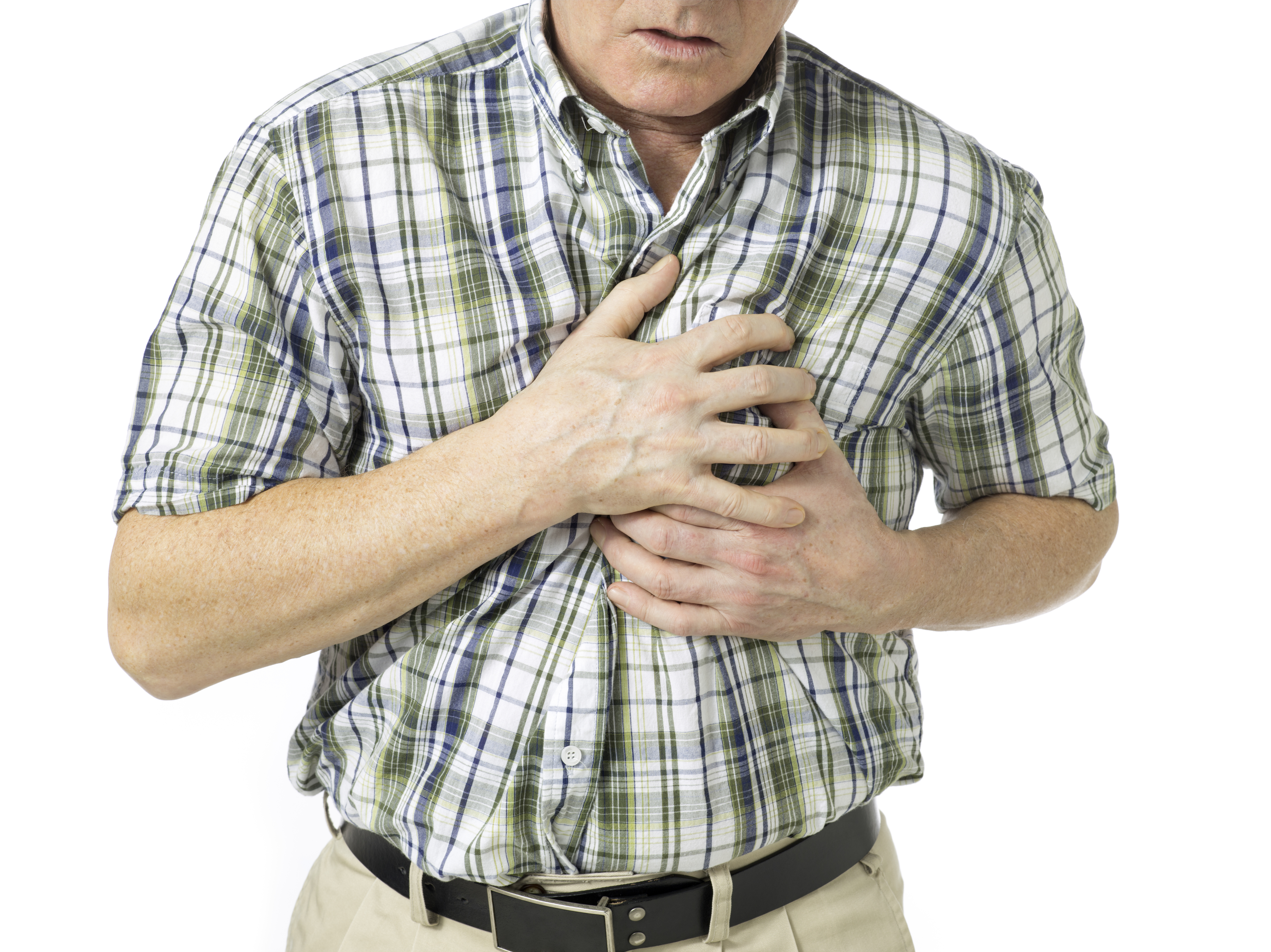




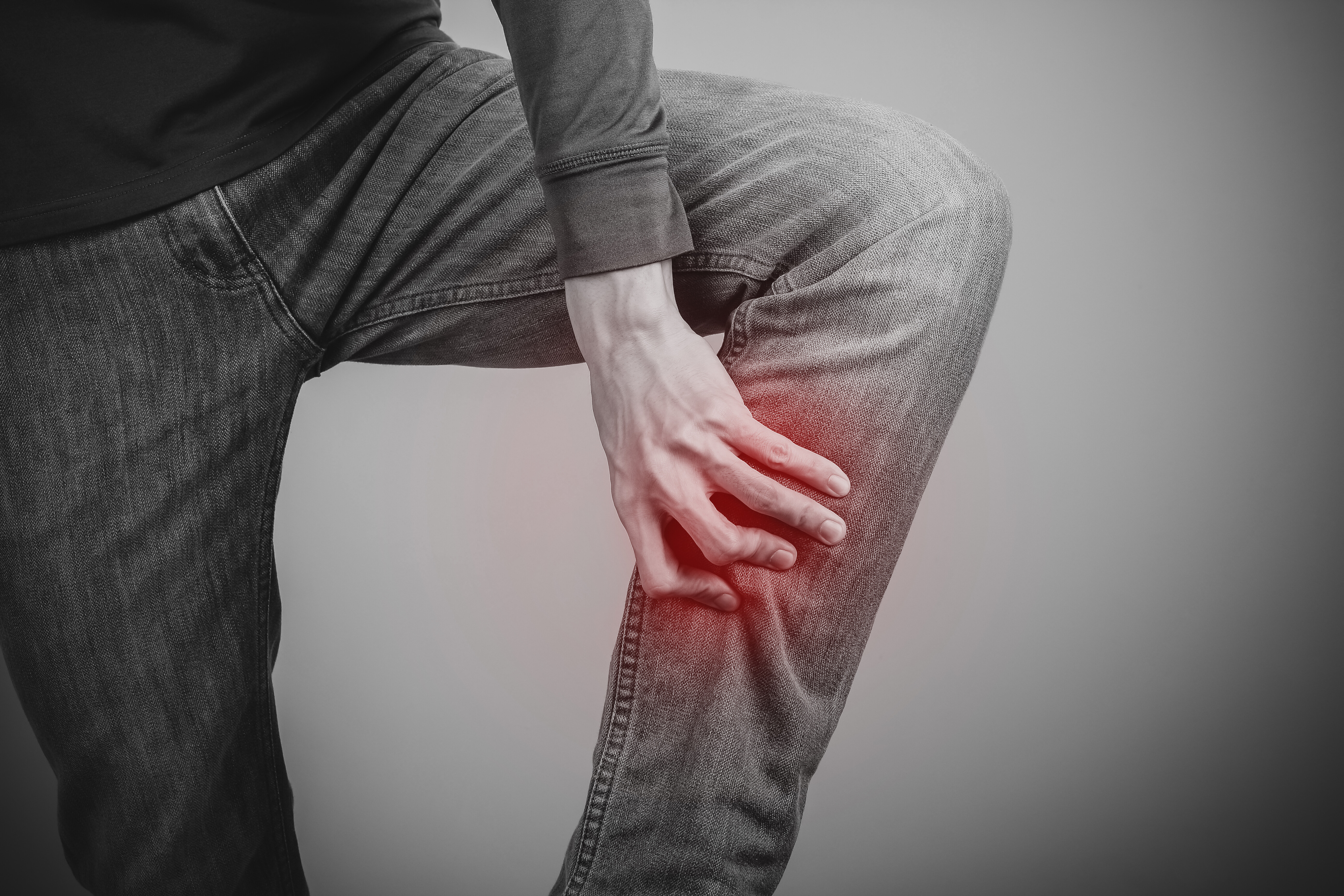

Can a DVT Cause Pain without Swelling or Redness?

We always hear how a deep vein thrombosis (DVT) can cause pain, swelling and/or redness in the affected area, which is commonly the calf.
The blood clot may also form in the upper leg or behind the knee. (more…)