Amy and Sheldon on “The Big Bang Theory” had a research collab that’s been a special interest of mine for years!
Some autistic people may identify with the characters of physicist Sheldon Cooper and neuroscientist Amy Farrah Fowler on “The Big Bang Theory” – characters coded for autism though not actually identified in the show as being on the Spectrum.
One of the hallmark traits of ASD is a lifelong propensity towards special interests: either a hyperfixation or “obsession” with a topic that may appeal to many neurotypicals such as dinosaurs.
The interest may also be highly specific or very unusual, such as the color indicators of license plate renewal stickers (yes, I met an autistic woman with this intense interest).
Not having viewed every episode of “Big Bang Theory,” I’m aware of only two Sheldon/Amy research collabs: symmetry and asymmetry in string theory; and the time lapse between a conscious decision to move a muscle, such as when picking up a spoon, and actually moving the muscle (picking up the spoon).
Well, that second one really resonated with me: The time it takes from when a conscious decision is made to move a muscle, to the time of the actual muscle contraction.
We’re talking milliseconds here, because nerve impulses travel 40 to 300 feet per second, or —depending on source of information — 210 to 360 feet per second or 156 to 270 miles per hour.
But the bigger thing here is the distance for which these speeds is confined: the length of the human body.
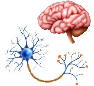
Dr. Fowler wanted to know the time lapse between the conscious decision to, for instance, wiggle a toe, to the time the muscles were contracting to move the toe. Damn! MY exact question beginning in my mid-teens!
Everything seems instantaneous, doesn’t it? There seems to be no time passage between when you “decide” to move an index finger and when the motor neurons innervate to contract the involved muscles!
But there IS time passage. It’s just imperceptible to us. Plus, a lot goes on during those microseconds.
Since early high school I’ve been fascinated by severed spinal cords.
It all began when the entire student body was treated to a movie in the school auditorium as part of the annual school celebration day, either in my ninth or tenth grade (decades before the Internet).
The movie was “The Other Side of the Mountain,” the true story of Olympic hopeful Jill Kinmont who suffered a ski injury that caused permanent quadriplegia.
Her spinal cord had been transected, but reattached. So I couldn’t understand why she was still paralyzed.
This bugged me. So next day after health class, I asked the teacher why, after everyone else fled the room at the sound of the bell.
While classmates at the all-girls school were interested in Rod Stewart and Peter Frampton, I was interested in severed spinal cords.
The teacher drew a stick figure and explained it in under a minute. I understood completely.
For readers who don’t understand why a person is permanently paralyzed despite the spinal cord being reattached, think of it this way:
Suppose the cord to your desk lamp is snipped in half. The light will go out.
Now suppose you take superglue and duct tape and reattach the severed ends. Will the light go back on? No.
That’s because, despite the reattachment, those ends are damaged. Once cells in the spinal cord are damaged, their function can’t be restored (even though mouse studies are promising).
So one day I asked a brother, who’s always been intrigued by human evolution (as is myself), if he had any ideas as to why damaged skin and bone cells easily grow back, while nerve cells don’t.
He gave a convincing answer: During human evolution, people were always getting skin injuries and breaking bones in their day-to-day survival.
Thus, skin and bone had plenty of “practice” to evolve to easily regenerate and repair themselves.
But severed spinal cords and brain injuries were very uncommon (no motor vehicle crashes, sports or workplace accidents, or falls from ladders during prehistoric times!), and hence, neurons never had enough practice to “learn” how to self-repair. Makes sense to me.
As a teen, I’d always think that by the time I was well into adulthood, there’d be a cure for severed spinal cords — some kind of gadget implanted between the severed ends, causing nerve impulses from the brain to “jump” across the electronic bridge and be picked up on the other side and make their way to the muscles to innervate them.
Yet here we are, decades later, with no such development for humans in sight.
And no, exoskeletons that the patient wears, that move their legs for them, don’t count. This movement is generated by an external unit, not something inside their spinal cord.
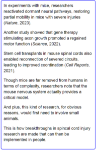
Mouse Studies Show Hope for Curing Spinal Cord Injury