

Can Popping Balloons at a Child’s Birthday Party Harm Hearing?

You may want to skip all the colorful balloon popping at your child’s next birthday party, as this can damage the hearing of little ears.
Certainly, good food and fun games are all that the partygoers need to have a great time. (more…)

How to Get a Dog to Do HIIT While YOU Take It Easy!

You don’t have to do HIIT just because you’re “making” your dog do it!
There’s a way to take it quite easy while you get your dog to do high intensity interval training. (more…)

Why Do Some People Always Smile when Talking Even if It’s Bad?

Do you know someone who, no matter what the topic of conversation, can’t quit smiling when talking?
This doesn’t refer to the smiling between two good friends who haven’t seen each other for awhile or someone telling a funny story.
This article refers to two kinds of “smile while talking” people:
#1 Someone who has a continuous smile on their face while talking to you, and this person may be someone you hardly see or frequently see, and you’ve noticed they’re like this with anyone they talk to.
So it has nothing to do with how likeable you are.
#2 Then there’s the news anchorman or anchorwoman, or some guest on a CNN or Fox News segment about the latest story.
Or, the “smile while talking” person may be an attorney, juror, reporter or someone somehow connected to a murder case and is giving some information.
But what’s there to smile about?
The striking thing is that the topic of what they’re speaking about is neither funny, amusing nor delightful.
Ever see a TV show called “Snapped” on the Oxygen network? These are documentaries of women who’ve committed murder.
Each episode features key people such as a detective, a juror, the killer’s mother, a friend of the deceased, etc.
Every so often, one of them has a continuous smile on their face as they’re talking about a MURDER.

One man, in law enforcement, was describing details about a decapitation — and all the while, had a smirk on his face!
There are guests on CNN or an ID Channel show who, while talking about some disastrous incident, such as a plane crash, mass shooting or serial rapes, is wearing an ongoing grin while explaining things and answering questions.
What is UP with this?
Look at the smiling woman below, taken from a crime docudrama called “Fear Thy Roommate.” What do you think she’s talking about?

The picture was taken as this woman, Tiffany, was describing how her mother Darlene’s roommate Angela would remove the shared home’s lightbulbs — to make the place dark — and hide her glasses.
Tiffany states that this “made my mother stumble around in the house and hurt herself.” Why is she grinning as she says this?
Eventually, Angela assaulted Darlene; Darlene called the police. Angela convinced the cops that she was the victim.
Darlene was taken away by the police. Below is how Tiffany looked as she said, “She got three days in jail.”

It gets worse. Tiffany is then talking about the time that Angela struck Darlene in the head with a hammer and then threatened her with a knife. Below is the image right as she’s describing this.

This is absolutely appalling. Not long after the incident, her terrified mother stabbed Angela to death after Angela began attacking her.
This is serious stuff, yet Tiffany is grinning all throughout.
And then there are the reporters who have a constant smile while talking about less tragic events.
But at the same time, these events are not the least bit funny, amusing or delightful, such as a traffic jam or local flooding.
Why do these people smirk?
One time on CNN was a guest who was commenting on Donald Trump’s presidential campaign.
The entire time she was talking, she was smiling — from ear to ear. It really looked ridiculous.
She had a very pretty face. Perhaps while growing up, she was always being told to smile — because of her attractive features, a la “You should smile more often; you have such a pretty face!”
Hearing this instruction often enough, she developed the habit of breaking into a fixed, ear-to-ear smile whenever she was talking about ANYTHING.
It just looked so scripted. And yes, it was ear to ear and very inappropriate for the topic of talk.
I’ve seen many reporters on TV wearing an ongoing smile (which is often more like a grin or smirk) while relaying a story about something that is nothing to smile about, such as a forest fire, tornado, street crimes, a segment on identity theft, etc.
Check out the image below of CNN anchor Abby Phillip.
The three other people have a serious, concerned facial expression, while Abby maintains a goofball smirk.
It wasn’t momentary; it was ongoing throughout the discussion; very inappropriate.

Woman Smiles Through Discussion of Sexual Misconduct Allegation
Below is an image of a panel discussion on Democrats staying silent about sexual misconduct allegations against Governor Mario Cuomo by a former aide.

Why is the woman on the bottom left smiling? Look at the other panelists. This was not a fluke second of time. This woman was grinning the entire time.
It’s easy to believe that someone in these individuals’ past kept harping on them to “wear a smile all the time.”
Perhaps as children they got punished for not smiling on command.
And it isn’t just women who do this. I’ve seen plenty of men with a continuous smile plastered on their face while part of a serious discussion or debate.
Smiling while talking of a heartwarming story is one thing, such as a child whose lemonade stand near a construction site made a killing.
But as mentioned, this refers to people who can’t stop smiling while talking about murders, violent crimes, racism, ISIS, the latest controversial subjects, bad weather, political scandals – you name it.

Some people wear a perpetual smile just like this, even when they’re on court TV and listening to the judge describe their case — even if it’s a very distressing case. WTF! Shutterstock/Tanit
I have an adult nephew who did this last time I saw him — but the conversation wasn’t about anything amusing; rather, just run-of-the-mill catch-up stuff, as I hadn’t seen him for a few years.
The nonstop grinning was annoying, I’m sorry to say. He was doing this with everybody, so it wasn’t an issue of him being ecstatic to chat with me (I’m not THAT special!).
The Problem with Smile Talkers
This habit can put listeners, in a face-to-face situation, ill at ease.
If a person is shy, introverted or otherwise socially awkward, they may feel pressured to smile back or maintain a perpetual smile themselves while the talking person keeps smiling away.
This is very unnatural and awkward to such a listener who’s already self-conscious enough as it is in social situations.
If this describes you, you’ll feel liberated if you wear your natural face.
Do not let someone else’s relentless habit dictate how to move the muscles of your face.
If something’s funny, then by golly, crack a smile. If nothing is funny, do not feel pressured to be untrue to yourself.
 Lorra Garrick has been covering medical, fitness and cybersecurity topics for many years, having written thousands of articles for print magazines and websites, including as a ghostwriter. She’s also a former ACE-certified personal trainer.
.
Top image: Shutterstock/UfaBizPhoto

Spinal Stenosis: Should Trying to Stand Straight Be Avoided?

As spinal stenosis progresses, the patient gets increasingly hunched over when walking.
“Spinal stenosis is a common problem that develops in the spine as we age,” says Dr. Nick Jain, MD, a board certified and fellowship trained orthopedic spine surgeon at DISC Sports & Spine Center in Newport Beach, CA.
“Oftentimes, we notice, as people with spinal stenosis start to hunch forward or their neck begins to tip forward.
“This typically occurs in the neck and low back as flexion (leaning forward) actually relieves spinal stenosis and provides more room for the spinal canal, and the body and nerves tend to prefer being in these positions when stenosis exists.”
Wouldn’t repeatedly standing straight help reverse this situation or at least make erect posture more tolerable?
“Stand up straight!” my mother has often told my father, who has spinal stenosis.
It’s not uncommon to see senior age people walking with the distinct hunching over of spinal stenosis (narrowing of the spinal canal through which the spinal cord fits).
Hunching over is called lumbar flexion and relieves the pain, which is why patients do this.
There is actually nothing mechanical blocking their ability to straighten out more.
However, this doesn’t mean in severe cases that they can straighten out all the way and stand perfectly vertical.
In my father’s case he can straighten to nearly vertical but claims that this position is intolerable. But when he lies on his back in bed, his back is perfectly straight.
“Extension (leaning back) and trying to stand very erect with good posture can actually compress the nerves and be more painful for people with spinal stenosis,” continues Dr. Jain.
Walking straight for some patients is so painful that they maintain a marked slump while walking even short distances.
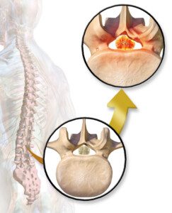
Spinal stenosis. Blausen.com staff/Bruce Blausen CC BY 3.0 creativecommons.org/licenses/by/3.0/Wikimedia Commons
From an intuitive standpoint, it seems as though an effective exercise would be to stand in one spot and very briefly straighten (extend), to retrain the back for good posture and strengthen the lumbar area.
It seems logical that over time, the duration of extension would increase due to a higher tolerance of this position that would come with time.
But what seems logical or intuitive isn’t always the reality.
“So, in short, for some people with spinal stenosis, trying to stand up straight probably should be avoided if it leads to increased pain,” says Dr. Jain.
“Forcing an erect posture can be dangerous for someone with spinal stenosis in the neck or low back, as it can actually kink the spinal cord or the nerves passing through the spinal canal — and should generally be avoided, as it would be unlikely to remodel the spine.”
“However, some people are hunched forward due to a spinal deformity that may be fixed or mobile.
“For people with these issues, core strengthening exercises and trying to maintain an erect posture may help eliminate pain and symptoms, as the body has a natural ‘cone of efficiency,’ and leaning or falling forward (i.e., bad posture) requires a lot of energy expenditure by the body and often induces pain.
“But people with spinal deformities without stenosis may benefit from exercises to improve their posture.”
Is it spinal stenosis or a spinal deformity?
Dr. Jain explains, “An evaluation by a professional can help distinguish between these two situations, as sometimes a small, minimally invasive treatment for spinal stenosis (such as an epidural injection or decompression surgery) can allow someone to stand up straighter and taller by alleviating the stenosis.
“However, people with spinal deformities often need surgical treatment to realign the spine and secure it in the proper place and alignment.
“Consult with an expert for your specific situation before forcing the neck or low back into a certain position, especially if that position causes discomfort, numbness or tingling.”
To put this succinctly, you can’t work out spinal stenosis.
 Dr. Jain specializes in adult degenerative spinal care and minimally invasive techniques and motion preservation surgeries. Recognizing that there’s no one-size-fits-all method to treatment, he analyzes each patient’s whole picture in determining what will yield the best possible outcome.
Lorra Garrick is a former personal trainer certified through the American Council on Exercise. At Bally Total Fitness she trained women and men of all ages for fat loss, muscle building, fitness and improved health.
.
Sources:
verywell.com/things-to-stop-doing-if-you-have-lumbar-spinal-stenosis-2696099
verywell.com/spinal-stenosis-try-this-exercise-first-2696098
rheumatologynetwork.com/articles/managing-degenerative-lumbar-spinal-stenosis

Will the Squeak in Your Hip Replacement Ever Go Away?

Your total hip replacement makes a squeaky sound with every step. Will the squeaking eventually go away or are you stuck with it? What about a revision surgery?

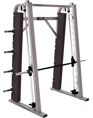
Smith Machine Bar in an Incline for Pull-Ups: What This Means
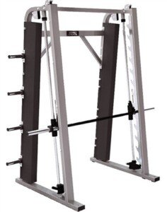
If you have a workout program that suggests “Smith machine bar in an incline” for women unable to do pull-ups, and you don’t know what this means, then here is the explanation.
Pull-ups Using a Smith Machine at “Incline”
When I hear “Smith machine” and “pull-ups” in the same sentence, an image comes to mind:
• Setting the Smith bar high enough so that you can hang on it, legs bent so that feet are off the floor, and do pull-ups—feet never touching.
• For women who can’t do pull-ups but are almost there, it can mean letting your feet contact the floor at all times to subtract some body weight, or having them make contact for some of the time throughout the repetition.
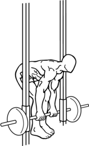
But when we toss “incline” into the picture, only one thing comes to mind: Setting the bar low enough so that your BODY is inclined, at an angle, as you pull up and lower.
In other words, a “Smith machine bar in an incline” is an odd descriptor for an inverted row or “modified pull-up.”
If the OP’s workout plan literally says “Smith machine bar in an incline,” then whomever wrote that must have been half-asleep at the time.
The OP thought that “incline” meant place an inclined bench under the Smith bar, sit in it, then pull up off the bench, then lower back into the bench.
I’d be extremely surprised if the workout plan really does call for this, as this would be a very inefficient way to do pull-ups and get strong enough for real pull-ups.
First off, it would be a hassle just to drag the bench over to the Smith machine every single time for pull-ups.
Now imagine sitting in an inclined bench, Smith bar over you, grabbing the bar and lifting yourself out of the bench. How cumbersome and inefficient can this possibly get?
“Incline” obviously refers to the inclined angle of one’s body during a reverse row or the so-called modified pull-up.
Smith Machine Pull-ups
• The bar can be set at any number of heights, depending on one’s strength and body height.
• The Smith machine makes for a very convenient reverse row apparatus and is often used for this purpose.
• Get under the bar, but make sure the height is high enough to allow full extension of your arms as you hang.
• Your body is straight out in front of you, but because the bar is higher than the floor, your body will be inclined.
• Your heels are on the floor, feet up.
• Keep your body straight and pull up. Your chest should come up under the bar.
• Keep your body as straight as a plank of wood throughout the set.
• If it’s too difficult to pull yourself all the way up, raise the bar.
• Raising the bar will make your body more upright but still “inclined.” But the move will be easier.
• This is the same move that you’ve probably seen many times being done using suspension straps.
To get strong enough for pull-ups, I recommend using the Smith machine bar while your body is 100 percent upright — but many women will need to build up to even this point.
Lorra Garrick is a former personal trainer certified through the American Council on Exercise. At Bally Total Fitness she trained women and men of all ages for fat loss, muscle building, fitness and improved health.

Should You Worry About Your 5-Year-Old Girl Being Too Tall?

If your tall 5-year-old daughter sees you worrying about her height, she’ll internalize this anxiety & learn to hate her body — and that can attract bullying.
Okay, it’s one thing if you’re concerned from a medical standpoint and are worried about a pituitary tumor because your five year old daughter is closing in on five feet tall.
But if she’s “a head” taller than her classmates, and healthy otherwise according to her pediatrician, you should not worry.
Otherwise, think of what the worry and anxiety will do to her self-image.
“If parents are hyper-focusing on any aspect of their daughter’s appearance and worrying aloud about it, that sends her an unfortunate message: Her appearance is more important than other aspects of who she is, and something about her appearance is not good enough,” says Patricia Celan, MD, a senior psychiatry resident at Dalhousie University in Canada.
“Society does enough of this – let her family show her otherwise.
“Don’t fixate on making comments such as, ‘You’re so tall,’ especially not in a negative or worried tone.”
A comment of “You’re getting so tall” may seem benign if spoken in an enthusiastic tone, but it can be a gateway to comments such as, “I sure hope you don’t get any taller,” or, “If you get any taller you’ll be bigger than all the boys on this block, and we don’t want that.”
Dr. Celan continues, “If your daughter seems to have a passion for sports, for arts or for learning in any particular subject, make sure you’re not diminishing the value of those things by letting her believe her world should revolve around her height.
“Try tailoring comments to be less about appearance and more about her interests and her personality, so she can develop into an emotionally healthy and fulfilled young woman rather than someone who is forever insecure about whether or not her appearance is good enough.”
Proof in the Pudding
There is a tall woman’s site that has a section where women could post their feelings about their height.
I never knew that so many tall women could actually be suicidal over this, but that’s what a striking number of comments say.
And even more of them literally weep over their height and blame it on all the bad things that have ever happened to them such as romantic relationships gone bad and childhood bullying.
They slouch, avoid heeled shoes like the plague and are constantly tortured by self-consciousness.
After reading their posts, you’d think they were cursed with giant brown hairy warts all over their face and fungus growing out of their head.
Instead, many of these women are only 5-11 and even 5-10! I say “only” because many of the posters with a more positive attitude are 6-2 and 6-3!
Parents Worrying About Young Daughter’s Height: Dangerous!
So your five-year-old girl is growing super fast or maybe has always been very tall for her age, towering over her kindergarten classmates and even being taller than older neighborhood children.
Sometimes she’s mistaken for a seven- or eight-year-old. Does this upset you?
Damn, stop it.
Ever see the commercials for St. Jude’s Children’s Research Hospital? Maybe you should pull these up on YouTube and watch them for awhile.
I’m pretty sure that every one of those parents in these commercials would trade their child’s cancer for a very long — and healthy — body.
Imagine learning your very tall five-year-old daughter’s leg pain is from bone cancer and that the leg needs to be amputated.

Bill Branson /ancer.gov
Hmmm, suddenly, her height won’t matter to you so much.
The women on the tall women’s site, I have to believe, had mothers who continually showed languish over their height growing up.
These women, as little girls, internalized this worry and unhappiness over their height and grew up to bear this cross.
It reminds me of an article I wrote for a magazine years ago about what makes a child so fearful of the dentist – even though they’ve never experienced any pain from a dentist.
The dentist I interviewed told me the cause was the parents. “The child sees the parents’ own anxiety over the dentist and they learn to fear the dentist. If Mom is afraid, then I should be afraid.”
It’s the same concept with height: “If Mummy is worried I’m getting so tall, then it must be a really bad thing.”
So the little girl learns – from her own mother (and maybe even Dad) – that being tall is something awful. The foundation has been laid; the trajectory for her growing-up years has been set.
Everywhere she goes she will feel mercilessly self-conscious about her height, and this self-loathing will attract bullies like a light drawing moths.
If your daughter is on course for being a very tall woman, then embrace it. It is just SO COOL when a woman is very tall!

The other day I saw a woman in line at McDonald’s – she must have been close to six feet.
Her husband must have been close to 6-4. He was holding their preschool daughter.
It’s a pretty good bet that the girl’s final height will be well over six feet, and that all through school, she’ll tower over her classmates. Lucky her!
If parents embrace their tall daughter’s height, this will set a very positive, rewarding trajectory. Quit your bellyaching and just be thankful your child is healthy!
Source: tallwomen.org
 Dr. Celan is a post-graduate trainee in psychiatry, working in diagnosing and treating patients with psychiatric conditions. She is passionate about psychotherapy, especially in trauma, anxiety and depression.
Lorra Garrick has been covering medical, fitness and cybersecurity topics for many years, having written thousands of articles for print magazines and websites, including as a ghostwriter. She’s also a former ACE-certified personal trainer.
.
Top image: Shutterstock/luckyraccoon

Guidelines for Jogging after Total Knee Replacement Surgery

Are you now fully recovered from your total knee replacement surgery and are interested in taking up jogging for aerobic exercise?
There is a strong likelihood that you, as a total knee replacement patient, were not a runner or jogger in the few years prior to the surgery.
By the time a TKR is needed, the joint is bone on bone or almost such, and the patient is suffering from considerable pain; even slow walking is very painful.
Perhaps many years ago you were into jogging, and now you want to get back into it.
Or maybe you’ve never been a jogger, much less runner, and are now considering taking up this form of cardio exercise.
However, as a total knee replacement patient, running—and even jogging—is off-limits to you.
Why Total Knee Replacement Patients Should Never Run or Even Jog for Exercise

Credit: Dave Haygarth
“I would never recommend that anyone (previous runner or not) who has had a TKR take up running, unless it is to run from someone for your life,” says Barbara Bergin, MD, board certified orthopedic surgeon at and co-founder of Texas Orthopedics, Sports & Rehabilitation Associates.
Dr. Bergin explains, “Many surgeons who do TKR’s don’t like to give patients any doubts regarding their knee replacements, so they often tell patients they can do anything they want to after surgery, like run, play sports and ski.
“If they tell you that you can’t do these things, patients often wonder why they would want to have the surgery if they can’t resume the things they used to do or the things they think they might like to do.
“They will have doubts about the integrity of the operation and the skill of the surgeon. So the surgeon just says ‘yes.’
“As a surgeon who used to do total joint replacements, I can tell you that I always told my patients ‘no.’
“And here’s why. You have only one chance to have one total joint (per joint). They last about 20 years before they loosen and wear out.
“This stat includes all comers, from 45-year-old, morbidly obese patients to 100 lb., 80-year-old ladies.
“If you have your total knee replacement when you’re 60, and it wears out when you’re 80, you are now faced with a revision.
“Revision joint replacement is a salvage operation. It is more complicated. More bone must be removed. You are more likely to have a fracture or an infection. You are more likely to be dissatisfied.
“Your surgeon assumes, if you’re older, that you aren’t going to go out and tear it up, so they say ‘yes.’
“But younger patients are more likely to get more active. This is why they are more likely to wear the total knee replacement out sooner and are more likely to be dissatisfied with the result.”
- The artificial joint includes polyethylene bearings.
- Running would place tremendous stress on these components which could lead to a disastrous failure of the polyethylene material.
Exercises that a TKR Patient Can Do
Dr. Bergin explains, “Take care of your total knee or hip replacement like it was your child. Don’t do high impact exercises. Don’t do squats. Don’t play sports.
“What can you do? Walk, swim, cycle, play golf, travel and just live without pain. That’s the actual reason to do a total knee replacement: to decrease pain.
“It’s not to allow you to do more activities, unless it’s just the activities of normal living.”
Don’t even think about skiing or racquetball, though light tennis is approved by the American Academy of Orthopedic Surgeons.
Yoga is also permitted, but do not sit on your heels and avoid squat-like poses or positions that involve considerable knee bending (e.g., child’s pose). There are plenty of yoga poses that are safe for an artificial knee joint.
“And a word to the wise,” adds Dr. Bergin. “Patients who are overweight often think that they need that knee replacement in order to exercise and lose weight.
“Warning: Studies show that pretty much no one loses weight after having a total joint replacement. And that’s because weight loss occurs in the kitchen and not out on the running track.”
Walk, Don’t Run, with a Total Knee Replacement

Shutterstock/ Dmytro Zinkevych
Proper biomechanics when walking are crucial for those with a TKR. Though walking outdoors is better for the joints than is using a treadmill, the treadmill is often a more convenient option.
Once you are completely recovered from your TKR, you should not hold onto the treadmill, as this will disrupt the body’s natural gait.
Or, to get a bit jargony, it will mess up the kinetic chain. This is the last thing a knee joint – artificial or natural – needs.
Holding on will force your body to assume improper posture while walking. There is no reason for continuously holding onto the treadmill and making the holding-on part of the workout.
Hold on while sipping water or taking a heart rate check, but then walk with your arms swinging naturally, the way the body is supposed to move.
If your knee hurts, this will not be corrected by holding onto the treadmill.
If you feel you’ll lose balance without holding on, then you’re going too fast, even if you think the speed is slow. It can always be slower.
Walking on a treadmill without holding on encourages good posture and proper knee movement because your body is forced to balance and walk correctly—the way nature intended it to.
The total knee replacement patient does not need to go running to get an effective cardiovascular workout.

Dr. Bergin is a general orthopedist, surgically and conservatively treating all manner of bone and joint conditions. She enjoys educating patients so they can emerge stronger than they were before their orthopedic injury or surgery.
Lorra Garrick has been covering medical, fitness and cybersecurity topics for many years, having written thousands of articles for print magazines and websites, including as a ghostwriter. She’s also a former ACE-certified personal trainer.
.
Top image: Shutterstock/MilanMarkovic78
Source: orthoinfo.aaos.org/topic.cfm?topic=a00357

Why Do Some Kids Foil Abduction Attempts and Others Don’t ?

What’s unique about the child who’s able to foil an abduction attempt, vs. ones who never try?
Every once in a blue moon you hear about a child who wildly “fights off” an abductor and escapes, while most other victims obediently do everything they’re told as though paralyzed with fear. (more…)

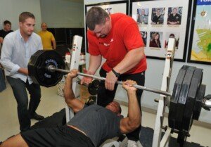
Weightlifting Exercises that Raise Blood Pressure the Most ?

Lifting weights will raise blood pressure, but there are exercises in particular that will really put your blood pressure through the roof.
Normal blood pressure is under 120/80. It’s not surprising that a very fit person with a clean diet has a resting blood pressure of around 100/60.
Any kind of resistance training will raise your blood pressure. Blood pressure can soar to 345/245 during very heavy lifts.
This was observed in a study (Journal of Hypertension Supplement, 1989).
The highest blood pressure increases during this study were caused by squats. Single-arm curls caused the lowest rises.

The back squat; raised blood pressure the most. Shutterstock/Photology1971
The report’s abstract does not list all the exercises that were monitored.
This will make one wonder if the deadlift and rack pull were part of this study, because these moves allow you to lift a lot more weight than does the squat.
However, we can definitely conclude that heavy deadlifts and heavy rack pulls will soar blood pressure — based on the premise that the back squat does.
All three exercises involve a barbell and mechanics that allow the body to move a LOT of weight.
Below are the exercises that allow the most amount of weight to be moved.
• Back squat

Shutterstock/MilanMarkovic78
• Front squat

Shutterstock/baranq
• Deadlift

Freepik.com
• Rack pull
Everkinetic, Creative Commons
• Olympic style lifts (e.g., clean & jerk)

Shutterstock/Microgen
• Bench press and leg press
Additional Factors Influence Blood Pressure
• Length of static holds
• Length of the set (e.g., 3 RM vs. 6 RM).
Baseline blood pressure influences how high BP gets when doing any lifting moves.
So if your baseline tends to be 90-something over 55 to 60, then when you deadlift a one RM, it won’t skyrocket as high as it would if your baseline were in the 120/70 range.
Make sure you get that: The BP will jump AS MUCH during a super heavy lift – whether your baseline is very low or average, but the final number won’t be as high if your baseline is very low.
In short, what gets tacked on during the lift is the same, but if it’s getting tacked onto a lower baseline BP, then the final number won’t be as high as it would if your baseline were higher.
Never hold your breath when lifting. Never. “This causes the intracranial, middle ear and chest pressures to go up,” says Dr. Beatty.
For big lifts with mega weight, this can put the athlete at risk for an intracerebral hemorrhage, especially if other risk factors are present such as a pre-existing brain aneurysm (which usually doesn’t cause symptoms and can exist without the person ever knowing).
Exhale on the lift and inhale on the release.
Though blood pressure will spike dramatically during heavy lifts, the long-term effect of weightlifting is a lower baseline BP.


