There IS a way to detect and diagnose melanoma when it’s in a very early stage — and hence, highly curable with an excellent 10-year survival rate.
Just how early can melanoma be detected? First of all, some exciting news:
The chief reason why melanoma cells are so resistant to chemo has been discovered by researchers at UC Irvine’s Chao Family Comprehensive Cancer Center.
However, researchers are a long way off from applying this discovery to melanoma treatment. This is why very early detection of melanoma is crucial.
Those who fear this skin cancer and/or are at elevated risk need to know about a technology called serial digital dermoscopy.
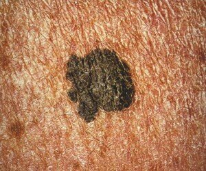
Malignant lesions can be missed by the layperson during monthly skin checks and even by a dermatologist, simply because the naked eye cannot always detect changes.
“Monitoring your moles for change or irregular features is one of the best ways to detect melanoma skin cancer early,” says Adam J. Mamelak, MD, a board certified dermatologist and founder of Sanova Dermatology in Austin, TX.
“In the doctor’s office, dermatologists often use handheld microscopes called dermatoscopes to evaluate the features and patterns of a mole or pigmented lesion on the skin.
“Just like the A, B, C, D, E features of atypical moles that we look for with the naked eye, there are a number of characteristics that we look at with the dermatoscope to determine if a spot on the skin is benign or malignant.
“Newer dermatoscopes allow you to connect a phone or camera to the magnifying lens and take pictures of the dermatoscopic image.
“Just as we look for changes in a mole with the naked eye, serial dermatoscopic images can be compared to make sure no irregular features are visible or become visible over time.
“DIgital dermoscopy, just like high resolution digital photography, has become a useful tool in the dermatology clinic.”
Serial digital dermoscopy
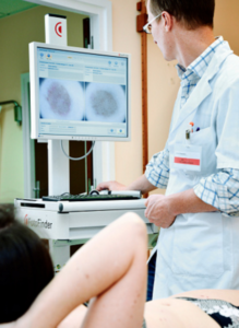
A dermatologist will examine your skin and then select the moles for the serial dermoscopy.
In my case, I requested that the moles on my back — even though typical — get imaged since I can’t easily inspect them.
Selected moles were indicated with a marker so that the nurse knew which ones to image.
Soon after, the dermatologist sat with me at the computer and reviewed the magnified images.
Serial digital dermoscopy involves comparing the images to a database of images of normal and melanoma moles.
Some computer systems will rate the images according to a color spectrum.
Other computer systems don’t do any rating, and instead, dermatologists use their naked eye to inspect the blown-up images on the computer screen for any signs of abnormalities.
When melanoma is caught very early, the 10-year survival rate is 99 percent.
 Dr. Mamelak