An ultrasound takes “pictures” of your heart.
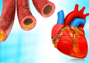
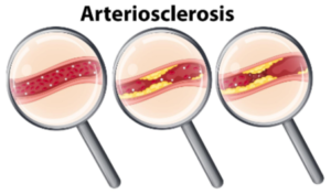
The difference between hard calcified plaque and soft plaque in the coronary arteries is that the hard buildup is much more stable.
The soft plaque, which is the dangerous type, can spontaneously fragment and cause a cascade of events that leads to a plug in the coronary vessel, causing a heart attack.
An ultrasound of the heart – also called echocardiogram – does not emit radiation.
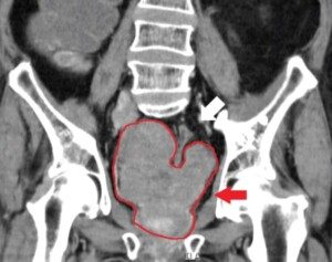
The CT scanner, on the other hand, emits radiation, whether the imaging is done for a calcium score or to detect soft plaque (CT angiogram).
The presence of a lot of calcified plaque, though not unstable like the soft kind, is still a very worrisome situation.
Ultrasound of the Heart: Looking at Plaque
“Echo has become very sophisticated, and the ‘pictures’ are remarkable,” says Michael Fiocco, Chief of Open Heart Surgery at Union Memorial Hospital in Baltimore, Maryland, one of the nation’s top 50 heart hospitals.
“But remember, they are generated from the reflection of ultrasound waves.
“That being said, echo is excellent at identifying high density in the wall of blood vessels, which means there is calcium there, and calcium is deposited into atherosclerotic plaque.
“The ability to identify plaque is excellent for the aorta and larger vessels, and with severe calcification can even be seen in the coronary arteries.
“Plaque that has not yet calcified can be very difficult to see with ultrasound/echo.”
As mentioned, to see soft plaque, a CT angiogram is the imaging modality.
An ultrasound may be the first imaging test that is done for a patient whose cardiologist suspects serious coronary artery disease (or an issue with the heart’s valves or chambers).
But a CT angiogram (which uses contrast dye) is far more telling when it comes to the presence of soft unstable plaque buildup.
And of course, the CT scan without the dye is used to determine a calcium score.
However, the gold standard for looking at both soft and hard plaque in the coronary vessels is the catheter angiogram.
The challenge with this imaging modality is that it’s invasive and brings with it a risk of stroke and heart attack, though that risk is small.
In addition to having the potential to give a cardiologist a good idea of the health status of a patient’s heart vessels, an ultrasound also yields other information such as structure, dimensions and function of the heart and its valves.

Dr. Fiocco specializes in treating artery disease, valvular disease and aortic aneurysm. His heart care expertise has earned him recognition by Baltimore Magazine as a Top Doctor in 2010, 2011, 2013, 2016 and 2017.
 Lorra Garrick has been covering medical, fitness and cybersecurity topics for many years, having written thousands of articles for print magazines and websites, including as a ghostwriter. She’s also a former ACE-certified personal trainer.
.


