
Here’s a simple explanation of what exactly “early detection” of melanoma means.
Melanoma is actually highly curable when caught early.
The problem is that the opportunities for an early detection are often passed over!
What Is Early Melanoma Detection?
From a medical standpoint, it’s the discovery of a melanoma in-situ or at stage 0.
From a layperson’s standpoint, it can be, but is not necessarily, the discovery of a spot on the skin that makes the person think, “What is this? I’ve never seen this before.”
And then at that point – not two months later – the person books an appointment with a dermatologist.
Another layperson standpoint is upon the first time they notice that a mole is changing, they make an appointment with a dermatologist (note: not the primary care physician), but again … the melanoma may already be beyond an early stage at this point.
This is why it’s crucial to check your moles and non-mole areas EVERY MONTH!
Very early detection of melanoma can be accomplished with groundbreaking technology called serial digital dermoscopy (more on this coming up).
What Is Early Melanoma? Stage 0 Explained
The epidermis is the top or outermost layer of skin. A stage 0 tumor is confined to the epidermis and is called in-situ.
Another component of very early melanoma is that its thickness is less than one millimeter.
Another indicator of earliness is what’s called a low mitotic rate (rate of cancer cell division).
Of course, a person can’t determine any of these features by looking at the lesion with naked eyes or even with a dermatoscope.
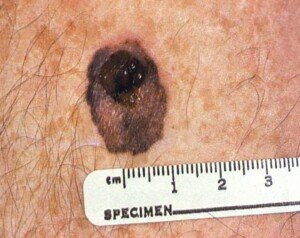
Early detection of melanoma does not always correlate to the size of the tumor, in that a tumor that’s two millimeters in diameter may already have penetrated the second later of skin (dermis), while a tumor that’s three millimeters in diameter may still be at stage zero (confined to the epidermis).
Early detection of melanoma is most likely accomplished by:
• Skin self-exams every month
• Clinical skin exams once a year (but nothing’s stopping you from having this done once every six months).
• Aggressive clinical exams if one has been diagnosed with dysplastic nevi (big and suspiciously shaped moles).
• Serial digital dermoscopy once a year
Serial Digital Dermoscopy (aka Mole Mapping)
• Every year your moles are photographed.
• The images are put in a computer and compared to a database of melanoma to determine a score for melanoma suspicion.
• The computer does not diagnose. It only yields a numerical or color rating of suspicion index.
• When in doubt, take it out. Suspicious moles should be removed and biopsied, even if the doctor says something like, “It looks benign.”
Listen to your gut. A good doctor will say, “It looks benign, but I’ll take it out and send it to the path lab if you don’t feel comfortable leaving it in.”
• The second time you undergo SDD, your updated images will then be compared to the previous images. The computer, as well as the doctor, can detect changes over time in a mole.
• The images are significantly magnified and enlarged on a computer screen and can be placed side by side for naked eye comparison.
Early detection of melanoma means a 10 year survival rate for stage 0 of 99 percent.
Mole mapping technology brings great peace of mind.


