The earlier that melanoma is caught, the more likely you can be cured from this vicious cancer.
Most people are not aware of a groundbreaking technology that detects melanoma far earlier than any clinical exam can.
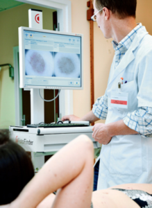
The technology is called digital dermoscopy, but this is a broad term, as this can refer to merely a hand-held device called a dermoscope that gives the physician a magnified view of moles.
A more in-depth version of “digital dermoscopy” involves a computer software program and serial photography of the patient’s moles.
Photos of moles, taken over a period of time, are taken, and then analyzed by the computer program to see if 1) they have endured changes over time, and 2) they have characteristics that, according to the computer’s database, resemble melanoma.
Digital dermoscopy with serial photography (around for about 10 years) is often referred to as “mole mapping,” though “mole mapping” is a broad term as well, and can simply refer to the patient’s crude drawing of his moles on a sketch pad.
I asked Dr. Theresa Pacheco, a dermatologist at the University of Colorado Hospital, about digital dermoscopy, aka mole mapping. UCH offers full-service digital dermoscopy including the computer software.
This technology is a godsend for people with many moles, and moles located in difficult-to-inspect areas like the back.
Elective removal of moles at increased risk for developing melanoma may make sense if the patient has only a handful of such moles.
But some people have literally hundreds of moles that require vigilant monitoring due to previous melanoma diagnosis, and/or nature of the moles (e.g., dysplastic).
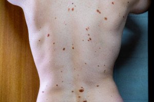
Many atypical moles. Shutterstock/Mikel Ugarte Gil
How effective is digital dermoscopy at viewing obscure areas like the scalp and genitals?
Dr. Pacheco: You can shave the hair or part the hair and still use digital dermoscopy to examine the lesion.
Basically, how does the computer software “detect” that a mole looks suspicious?
Dr. Pacheco: The computer program utilizes certain variables like ‘color,’ ‘symmetry,’ etc., that it utilizes to compute a score… our mole mapper rates each lesion as red (abnormal), yellow (quasi normal) or white (normal)…. but it is really up to the physician to determine whether a biopsy is warranted.
Does the device look at only moles (the moles that the dermatologist has determined for mapping)?
Dr. Pacheco: Yes.
Does the instrument also have the capability of detecting early melanomas in non-mole areas of skin?
Dr. Pacheco: No.
Basically how does digital dermoscopy work?
Dr. Pacheco: The mole mapper should be considered as a tool for use by a trained dermatologist — the dermatologist determines which moles to map….and ultimately which moles to biopsy to look under the microscope.
It is not possible (time) to map every square inch of the body.
We do take overview of body areas (trunk, arm, hand, etc.).
We know melanoma arises in existing nevi (but most nevi are normal) and equally in normal skin … so we focus on new moles, changing moles, symptoms associated with moles, etc. (“nevi” means moles).
Suppose a patient decides to have digital dermoscopy every month. Is there any medical impracticality to this?
I’d think that monthly digital dermoscopy would be superior to the patient’s own layman’s naked eye inspections, especially since the patient can’t possibly get an accurate view of his back; and that digital dermoscopy would take substantial guesswork out of self-exams.
Dr. Pacheco: Sure… but I usually recommend a baseline and then in six months. If moles are stable, I have them come back in a year.
Mole mapping has been hailed as highly effective for people with many moles/dysplastic nevi and a history of melanoma. However, what is your take on someone without “high risk” skin having digital dermoscopy done, say, once every six months, just for 1) peace of mind, and 2) superior detection over the layman’s naked eye, especially when it comes to viewing the back?
Dr. Pacheco: No system or doctor is perfect – so I am fine with patients requesting mole mapping. It does lessen the anxiety.
Although – usually after a couple normal/stable mole mapping sessions – they are fine with coming less often and letting me dictate control of their skin examination.
Can digital dermoscopy detect changes in a pre-existing mole before those changes can be detected with the most scrutinizing naked eye?
Dr. Pacheco: Yes.
In that case, what percentage of such early detection (when mole is biopsied and declared melanoma) are stage 0 or in situ? (very early melanoma)
Dr. Pacheco: I am not aware of statistical reports.
Are you aware of any technology on the horizon that will be able to scan the entire skin surface and thus detect malignant changes in their most rudimentary stages in non-mole areas?
No.
So if you are interested in the earliest detection of melanoma and are concerned about your risk factors, inquire about digital dermoscopy with serial photography to your dermatologist.