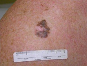
If you have a severely atypical, irregular or dysplastic mole that’s showing signs of regression (new areas of milky-white color), this is very worrisome for melanoma.
A white “spot” in a mole isn’t always cancerous, and may be a harmless hair follicle.
It’s very important to closely monitor any areas of even a normal-looking mole that appear to be disappearing — or seemingly filling in with a dull white or the color of one’s baseline skin tone: in other words, regressing.
Have you been told you have dysplastic moles?
These moles tend to be larger than average, often larger than the diameter of a pencil eraser, and usually are asymmetrical, lopsided, “odd” or “funny” in appearance.
Regression in Moles: Can Be Normal, Can Mean Melanoma
“Regression is a phenomenon in which a mole appears to be fading or disappearing, often in an irregular manner,” says Kara Shah, MD, an adult and pediatric dermatologist and founder of Kenwood Dermatology in Cincinnati, OH.
“To the naked eye, regression typically appears as one or more areas within a mole that are lightening in color,” continues Dr. Shah.
“Complete loss of pigment in some areas may result in areas that appear milky white in color.
“Regression is an immune-mediated phenomenon that results from activation of a type of white blood cell called a T-lymphocyte.
“It is considered part of the body’s innate cancer-surveillance system.
“Although benign moles may sometimes gradually and uniformly fade as part of a phenomenon called a halo reaction, signs of regression in a mole that appears atypical are concerning for melanoma.

Halo nevus (mole)
“If such a mole is biopsied and the dermatopathologist notes that the mole is severely atypical or dysplastic, the areas of regression may represent an area of regressing cutaneous melanoma.”
If you have an irregular or odd looking mole that has recently begun showing signs of regression – you should promptly make an appointment with a dermatologist.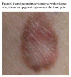
Do not settle for a naked-eye exam. The dermatologist should use a handheld lens (dermatoscope) to inspect the mole.
If the doctor says that the mole appears to be atypical or dysplastic, and that there’s signs of regression, it should be biopsied.
In fact, the development of new areas of white in even an otherwise normal-appearing mole may represent areas of regression suspicious for melanoma.
Malignant regression in any mole, for that matter, will appear in specific areas rather than causing a diffuse, evenly distributed regression.
When the entire mole seems to be fading evenly throughout, or diffuse fading – this “can be a sign of natural involution of normal melanocytic nevi, which is common in the aging population but can be selectively noted in younger persons and is generally a benign and harmless phenomenon,” says Dr. Shah.
Mole with Regression from Melanoma Is More Dangerous than Typical Melanoma
“If the T-lymphocytes have been mobilized to destroy melanoma cells as evidenced by areas of regression, it may make it more difficult to identify the melanoma on the skin, as some of the visual cues (e.g., brown or black areas within the melanoma) have been erased,” says Dr. Shah.
Consequently, diagnosis may be delayed.
What is a better description of an atypical mole with regression?
This would be “parts of it are disappearing” rather than “it’s fading.”