

If you struggle to get a good look at all the moles on your back when you do your skin cancer self-exams, there’s a solution to this problem.
It is called serial digital dermoscopy.
Though serial digital dermoscopy is still not a standard screening procedure for melanoma, it’s a pretty reliable way to follow hard-to-see moles on your back.
What is serial digital dermoscopy?
• A nurse circles with a marker the moles on your back.
• She photographs each one.
• Each image is entered into a computer database.
• Images are analyzed based on the database – analyzed with a color or numerical scoring system that ranges from a normal rating to a highly suspicious for melanoma rating.
• Your dermatologist will go through each image with you and its score. The images are magnified considerably on the computer screen.
• Every year you get the same moles photographed. Once there are past images for reference, the computer is able to compare the most recent images with the previous ones to detect any changes that are not visible to the naked eye.
• If you’ve been getting this melanoma screening procedure done for, say, five years, the doctor can pull up all five images of the same mole simultaneously for viewing.
You can also have other moles photographed; they don’t have to be located on your back.

Trying to get a good look at the moles on your back is difficult. If you have presbyopia (things are blurry up close) it’s impossible.
Viewing moles on your back (with mirrors, of course) can even cause anxiety, because you might discover a very dark spot that wasn’t there a month ago.
This happened to me. The spot appeared black, but I also suspected that it was a “hemorrhage.”
This is medical lingo for a tiny scab that’s shaped like a common mole. These stick to the skin for up to three weeks before falling off naturally. They can arise from scratching your back.
I rubbed my finger pad at the spot (never try to pick off or scratch off a mole or new growth with your fingernail).
I figured if the soft smooth pad of my fingertip could remove it, it must be a hemorrhage. And guess what: It came off.
I was able to catch it and view it in my palm under a magnifying glass. It was clearly a tiny scab.
The area of skin where it’d been looked perfectly normal.
When you inspect the moles on your back, you may be viewing benign skin barnacles called seborrheic keratoses – thinking these are moles.
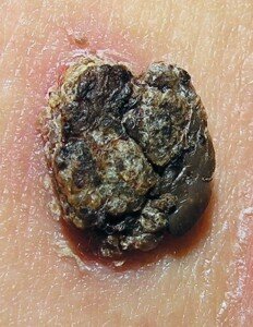
Up close seborrheic keratosis
These harmless growths can resemble melanoma but will NEVER turn into melanoma.
Seborrheic keratoses are not photographed for digital dermoscopy; only moles or lentigos are.
Lentigos are pigmented lesions that to the naked eye are often indistinguishable from moles.
My total out of pocket cost for serial digital dermoscopy is $250. This buys me incredible peace of mind because my presbyopic eyes just cannot clearly see the moles on my back.
Even if I had sharp close-up vision, I still would not be able to get my eyes close to the skin of my back like I can my arms, shoulders and face.
Insurance will not cover serial digital dermoscopy unless your dermatologist authorizes this.
If you have a large number of dysplastic nevi (atypical moles), your dermatologist may recommend serial digital dermoscopy.
If you have merely a number of normal common-looking moles, don’t bet on the recommendation.
However, the $250 is well worth it. That’s how much many women spend in just 30 days on cosmetics, manicures, pedicures, facials and hair care.
Men can easily drop $250 a month on beer, pizza, sporting event tickets, a lobster and steak dinner, a night at the movies and a few new CDs.
So don’t even think that the cost of serial digital dermoscopy is not worth it. CAN you get up close and personal with the moles on your back?


