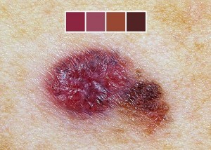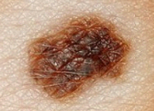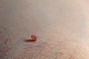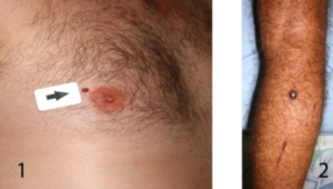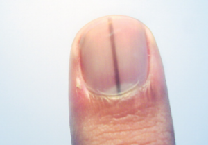
Only a biopsy has the final word on whether or not a worrisome mole contains melanoma cells or is entirely a melanoma.
That suspicious mole should be biopsied, even if your dermatologist says it “probably isn’t” melanoma after viewing it through a handheld lens known as a dermoscope or dermatoscope.
Though statistically, chances are in your favor that the questionable mole is benign, you should still get a biopsy.
“Whether you feel uneasy or your doctor is recommending a biopsy for a suspicious spot, it’s important to get the spot checked,” says Dr. Gretchen Frieling, MD, Triple Board Certified Boston Area Dermatopathologist.
“When you feel nervous about a spot, you can consult your skin specialist who will assess the spot and recommend a biopsy,” continues Dr. Frieling.
“If the doctor feels the spot may be something else, they can advise you accordingly. But when it comes to melanoma, the benefit of getting tested and finding the condition in time outweighs any risk of being overly cautious.”
Though melanoma has gotten a lot of media attention over the years, and coverage seems to be increasing, it’s still rare for moles to transform to melanoma.
However, about two-thirds of melanoma tumors arise in areas of skin where there was no pre-existing mole.
At Frontiers in Optics 2010 (Oct. 24-28), Duke University scientists presented a new technique for aiding dermatologists in helping to differentiate normal moles from melanomas, by using high-resolution snapshots of moles that appear suspicious.
About 25 percent of dermatologists use a dermatosope for routine checks of a patient’s moles.
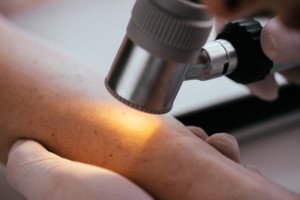
Dermatoscope. Shutterstock/LightField Studios
Certainly, you can imagine how long it would take for a routine skin inspection if the doctor used the dermatoscope to inspect every one of a patient’s 150 moles. Some people have even more moles.
On the other hand, some doctors don’t use this tool even for patients with a small number of moles.
It’s perfectly okay for the patient to request that the practicioner use this special magnifying device.
But this special lens is not a diagnostic tool. Though it enables the doctor to see things that he can’t with the naked eye, it’s still no match for a biopsy.
My doctor took me by surprise during one of my annual skin exams by saying, “You have a mole on your upper arm that gets my attention, and I’d like to get a biopsy on it.”
This was after she examined it with the dermatoscope. (The biopsy was done – entire mole removed).
She said a few times that it was “probably benign,” but you’ll want your doctor to always be ahead of the wave. My mole turned out to be benign.
In short, a mole that gets even only the attention of the patient should be biopsied. Trust your gut.
Cancerous changes can take place too deep within the mole to be seen with the handheld lens, though there are cases in which the physician not only can clearly see cancerous signs with the instrument, but with the naked eye as well.
However, there are times when even a biopsy nets disagreement among doctors.
So what about that study?
The Duke researchers proposed a two-photon microscopy technique that infuses a small quantity of energy into the two kinds of pigments that are found in skin: pheomelanin, and eumelanin.
The energy redistributes to yield high-resolution images of the pigments’ distributions, indicating possible melanoma or not.
“No one has been able to look at where different melanins are organized in skin,” says the lead researcher in the report. “This opens up a whole new pathway of looking for melanoma.”
At any rate, whenever you have a mole removed, even just for cosmetic purposes, insist upon a biopsy, just to play safe; a normal looking mole may end up being melanoma after all.

 Dr. Frieling’s website is