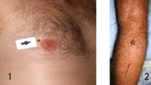
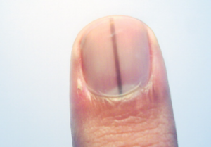
If your nail matrix was sent for biopsy due to a suspicion for subungual melanoma and it came back negative, you can be rest assured that you do not have this disease.
Unfortunately, there is no simple way to definitively diagnose melanoma of the nail matrix (known as subungual melanoma).
You may feel unsettled at the idea of having a portion of the top of your finger removed for the biopsy.
However, if this is what your dermatologist advises, there’s a good reason.
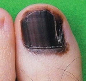
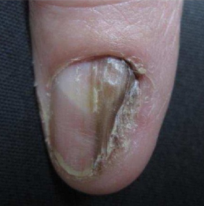
• Melanoma of the nail unit can clinically resemble a benign pigment.
• And a benign pigment can clinically resemble the cancer.
This cancer is as deadly as any other kind of melanoma if it’s allowed to metastasize.
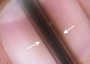
Even under a dermatoscope (a magnifying tool used by dermatologists), subungual melanoma cannot be diagnosed, though it can be strongly suspected.
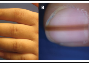
It may also have features consistent with a benign longitudinal melanonychia (pigment in a fingernail or toenail).
Reliability of Biopsy for Subungual Melanoma
“Biopsy is the gold standard and VERY reliable in diagnosing nail melanoma — provided the biopsy is done correctly,” says Adarsh Vijay Mudgil, MD, double board certified in dermatology and dermatopathology, and founder of Mudgil Dermatology in NY.
“The biopsy needs to capture the nail matrix (the most proximal part of the nail), which is where the nail is formed.”
This is what a layperson would call the “bottom” of the nail, or near the cuticle.
“A simple nail clipping won’t provide much diagnostic value,” says Dr. Mudgil.
This is because the tumor begins in the nail matrix (which is beneath the nail, hence the term subungual), not the actual “nail” that people trim, clip, chew or paint.

 Dr. Mudgil