

Blue arrows point to Hutchinson’s sign from melanoma shown under magnification. Copyright ©2018 Bhat et al
If you know what a Hutchinson’s sign is, perhaps you’re wondering just what is a micro-Hutchinson’s sign and how predictive it is of death by melanoma.
“Micro-Hutchinson’s sign is periungual pigmentation observed with dermascopy, which cannot be appreciated by the naked eye,” says Zain Husain, MD, FAAD, board certified dermatologist and fellowship-trained Mohs micrographic surgeon with New Jersey Dermatology & Aesthetics Center.
Micro-Hutchinson’s sign involves the same process as Hutchinson’s sign:
This is the expansion or proliferation of cancerous cells into the skin of the nail fold, and sometimes beyond that.
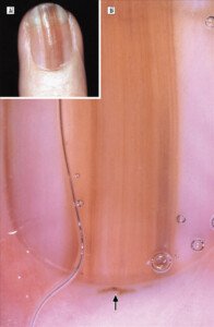
Micro-Hutchinson’s sign. Archives of Dermatology/ 2002;138(10)1327-1333
When this is detected by magnification via dermatoscope, but not visually with the naked eye, it’s deemed a micro-Hutchinson’s sign.
The apparent “spread” of pigment into the cuticle, nail fold and surrounding areas of a nail (the periungual area) is a very worrisome sign – even when it’s detected only upon magnification.
“It correlates with the initial radial growth of melanoma into the nail unit,” says Dr. Husain.
“Although it is a rare sign, its presence is highly suggestive of melanoma. A biopsy must be performed to establish diagnosis.”
 Dr. Husain has extensive training in advanced skin cancer surgery and reconstruction as well as cosmetic procedures including injectables, body contouring and laser surgery. He delivers lectures on dermatologic topics at national conferences.
 Lorra Garrick has been covering medical, fitness and cybersecurity topics for many years, having written thousands of articles for print magazines and websites, including as a ghostwriter. She’s also a former ACE-certified personal trainer.
.


