

If your dermatologist told you that a mole is too tiny to be biopsied, and to wait a little longer to see if it gets bigger, get a second opinion.
“Should physicians in the clinic assume a spot is too small for the pathologist to examine? No way!” says Jena Martin, MD, a board certified anatomic and dermatopathologist, trained at Mayo Clinic Pathology, having practiced in the Minneapolis area for over 10 years.
If you can SEE the mole, it’s not too small for a biopsy!
“We examine very small specimens in pathology; that’s why we use a microscope!” says Dr. Martin.
“If the dermatologist or clinical doctor takes a punch biopsy, we then cut in the tissue to prepare the biopsy for microscopic examination.
“The histology technicians cut the tissue to four micron thickness, so there is little chance we’d miss any cells. A micron is 0.001 millimeter.”
To put this very tiny size in a different perspective, a single red blood cell is seven microns in diameter. So you can see just how tiny that tissue sample is.
“When we look under the microscope at moles, it is true that we are looking for both pattern and the appearance of the cells,” explains Dr. Martin.
“Sometimes the pattern in very small moles is more difficult to interpret, but this does not mean these shouldn’t be biopsied. I have seen lots of benign (harmless) tiny moles and occasionally melanoma in a tiny spot.
“No other tumor is as small and as deadly as a melanoma. Dermatopathologists are mostly interested in the depth of the tumor — how deep does the melanoma invade below the skin surface?
“This is called the Breslow depth. We measure this in millimeters, and for perspective, one mm is considered a significant depth of invasion and is a cutoff for staging.
“Even melanomas that invade just more than 0.7 mm can be considered for sentinel lymph node biopsy when they are re-excised.
“A melanoma that is deeper than one mm is very deep. We measure invasive melanoma to a tenth of a millimeter; 0.1 mm invasion into the dermis.”
The dermis is the second layer of skin. The top layer is the epidermis.
“Many melanomas are not invasive and instead stay in the epidermis, called in situ melanoma.”
Biopsy of a Very Small Mole
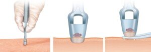
Punch biopsy. Shutterstock/ilusmedical
“Lots of factors go into a dermatologist’s decision to biopsy an atypical looking pigmented (dark) spot,” says Dr. Martin.
“Size is important, but it shouldn’t be the only factor in making this decision. Some clinicians prefer to biopsy moles that are over four mm in diameter so that patients are not over-treated.
“This is because we don’t really understand the biological behavior (meaning, can they become melanoma?) of very small atypical pigmented lesions. These could be early melanomas or they might not progress farther.
“However, even a smaller pigmented spot can be suspicious to either the dermatologist or the patient.
“The bottom line is if the patient is concerned about a new dark spot, they should ask their dermatologist to evaluate it and consider a biopsy.”
 Dr. Martin trained at Mayo Clinic Pathology and has been practicing for 20+ years.
 Lorra Garrick has been covering medical, fitness and cybersecurity topics for many years, having written thousands of articles for print magazines and websites, including as a ghostwriter. She’s also a former ACE-certified personal trainer.
.


