

It’s very true that an ultrasound can actually miss a DVT, but other tests are more definitive.
Staci Stringer’s DVT was missed by an ultrasound.
In September 2010, she experienced a deep vein thrombosis and pulmonary embolism.
When the DVT developed, Stringer was on birth control, but she also had lupus anticoagulant syndrome and rheumatoid/psoriatic arthritis.
Ultrasound Misses DVT
“It started as a pain in my leg, and after a week I went to the ER and had an ultrasound; they said it was nothing,” says Stringer on her site.
“The following Monday I was admitted to the ER with a PE,” which had caused chest pain.
“After a week or so the PE cut off blood flow to a portion of my lung which caused a pulmonary infarction.”
Another way of saying this is a “heart attack in the lung,” in that oxygen was cut off by the blood clots to the lung tissue, permanently damaging it.
Symptom Detail
“I started to feel the symptoms of a DVT in my right calf,” says Stringer’s account.
“It was swollen, extremely painful, hard to walk. I ignored the symptoms because I figured it was just arthritis pain.”
She continues: “I spoke with my general practitioner after a week and she told me to go to the ER for an ultrasound.”
Ultrasound is a standard diagnostic tool for DVTs.
“The technician thought he saw something but sent me home. I started to get a fever; my calf became so stiff I couldn’t walk.
“I walked up two flights of stairs to my apartment and was so out of breath I was grasping for air for 10 minutes.
“Next day I walked up the stairs to my office and I was so out of breath I fainted. I was admitted to the ER. After the CT scan they found a pulmonary embolism.”
How does an ultrasound miss a DVT?
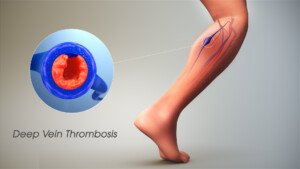
Scientific Animations, Creative Commons/BY-SA/Attribution-ShareAlike 4.0 International
“I regularly order a venous Doppler to rule out DVT,” says Reena Patel, MD, a board certified family medicine physician who treats patients at Garnet Health Urgent Care in NY.
“An ultrasound uses sound waves and compression [of the blood vessel wall] to see blood flow in the veins of an extremity and can find an obstruction.
“There is room for error. Specifically, human error can result in a false negative, so it’s important to see the patient as a whole.”
A probe is used and if it slides off the vessel wall, a false-negative finding can result.
“It’s important in my experience, if suspicion is high and Doppler is negative, to have a backup plan,” says Dr. Patel.
“I may order a D-dimer. This is a sensitive test and can be very helpful to back up your diagnosis and warrant further imaging.
“I consider their history, the current symptoms and calculate their risk using reliable scoring such as the Wells’ score [a numerical value derived from the patient’s answers to numerous questions].”
Additionally, if you believe an ultrasound has missed a DVT, you should ask about a color flow venous duplex scan.
This should be of the vessels close to your pelvis as well as extending down to the foot.
If the D-dimer blood test is positive, the protocol is to have the patient undergo a chest CT scan.
The younger the patient, the more likely that a positive D-dimer means a blood clot, since old age can cause a false-positive with the D-dimer.
What caused Stringer’s DVT?
“The doctors weren’t sure if it was my birth control, my arthritis flare that caused the DVT,” says Stringer, “but I later found out I have lupus anticoagulant syndrome. I’m on warfarin for life.”
 In addition to treating many chronic conditions,


