
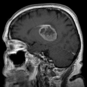
Did your MRI show a high grade glioma? Could it possibly be low grade?
How accurate is the MRI at telling a high grade from a low grade brain tumor?
A glioma type of brain tumor develops from astrocytes, which are star shaped (hence the prefix “astro”) glial cells that support nerve cells.
When the glial cells or astrocytes grow out of control (cancer), the mass is called an astrocytoma.
It may also be called a glioma.
A high grade glioma is aggressive, growing faster than does a low grade.
The way a brain tumor appears on an MRI – along with its location in the brain – can be very telling as to what kind of tumor it is.
High Grade Glioma vs. Low Grade on MRI
“An MRI can provide useful information to help diagnose types of brain tumors,” says Sumeer Sathi, MD, a neurosurgeon with NYU Langone Health, who treats brain tumors.
“What is shown on the MRI can closely resemble one type of brain tissue, and it can turn out to be a different type.
“The only definitive diagnosis can be made through obtaining a biopsy (brain tumor specimen) and sending it for pathology.”
 Dr. Sathi’s expertise includes spine surgery and treating brain tumors including metastasis, gliomas, meningiomas and acoustic neuromas using gamma knife radiosurgery.
 Lorra Garrick has been covering medical, fitness and cybersecurity topics for many years, having written thousands of articles for print magazines and websites, including as a ghostwriter. She’s also a former ACE-certified personal trainer.
,


