
Okay, we all know that “black and ugly” describes melanoma, and online images show melanoma to be evil-looking, dark, jagged, bumpy, the whole works.
But we must also consider that often, by the time a melanoma is photographed by a dermatologist, it’s at a later stage of development.
Who spots a tiny innocent-looking dot, says to their doctor, “I’ve never seen this before,” and the doctor decides to take a photo of it before excising it?
What more likely happens is that when what obviously appears to be a melanoma, with classic features, is presented to a dermatologist, the doctor THEN takes the photo. And it ends up online—looking mighty nasty.
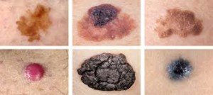
Note that the dark pink melanoma in the lower left is quite round, symmetrical and all the same color.
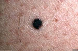
In actuality, not all melanomas—by the time they’re diagnosed—are scary looking or have any of the classic features such as jagged edges and bumpy portions.
This one here below is relatively symmetrical, and keep in mind that it’s magnified — which accentuates asymmetry in any lesion.
Melanoma Has to Start Somewhere
And it can start as a tiny sane-looking dot. A melanoma doesn’t go from nothing to a pencil-eraser-sized purple and black cobbled spot overnight.
“Not all melanomas begin as the typical funny looking or sinister lesion,” says Sharyn Laughlin, MD, board certified dermatologist and co-founder of the DermaEnvy Skincare ™ line of sun protection products and medical director of Laserderm, a pioneering laser skin surgery clinic in Ottawa, Ontario, Canada.
“Some start out as a very small, innocent looking, pale, pink or black spot or nodule that will over time change in size — a vertical growth rather than the typical spreading lesion.
“Some resemble a skin tag or a seborrheic keratosis. If the patient says this is new – take them at their word regardless of what it looks like and follow it with photographs or biopsy it immediately.
“This is where the experienced dermatologist exercises intuition – call it a ‘gut feeling’ and usually does the right thing.
“My philosophy is simply to biopsy it if any uncertainty persists after looking at the lesion and using my dermatoscope to see it under magnification.
“Then neither you nor the patient need lose sleep or worry. The pathologist gives you the definitive diagnosis.
“The downside — the patient ends up with a small scar, which in my hands using my CO2 laser to deal with the excision base, is practically invisible.”
A melanoma’s only distinguishing feature may simply be an increase in size over several months, even though the color, symmetry and border remain the same. When it doubt, have it checked out.
 In practice for 30+ years, Dr. Laughlin has been lead or co-investigator in many research trials and an innovator in developing new laser technology. dermaenvy.com.
 Lorra Garrick has been covering medical, fitness and cybersecurity topics for many years, having written thousands of articles for print magazines and websites, including as a ghostwriter. She’s also a former ACE-certified personal trainer.
.


