
Which is it?
Has a doctor recently told you “It’s a seborrheic keratosis” and you’re still scared it’s a melanoma?
Here’s what you should do.
A melanoma can sometimes be mistaken for a seborrheic keratosis which is a benign skin barnacle.
“There are MANY pigmented skin lesions and many can look alike,” says Jennifer Gordon, MD, who is board certified by the American Board of Dermatology; she practices at Westlake Dermatology located in Austin, Texas.
“These include moles, freckles, seborrheic keratoses, dermatofibromas, basal cell and squamous cell carcinomas, melanomas and many others,” continues Dr. Gordon.
Melanoma vs. Seborrheic Keratosis
“When these lesions are ‘text book,’ they can be fairly easily deciphered by the trained eye of a dermatologist; however, as we know, skin lesions don’t always read the textbook,” explains Dr. Gordon.
“True pigment can occur in many of these lesions, but also some of these lesions grow with a brown discoloration and thickening, which can make detection of underlying pigment in the actual skin difficult.”
How does a melanoma get past a dermatologist’s trained eye to recognize the distinctive pigment network of a melanoma?
And to this question of mine, Dr. Gordon replies, “There is no ‘distinctive pigment network’ of a melanoma. There are many red flags that a pigmented lesion might be a melanoma or atypical, but there is no perfect formula.
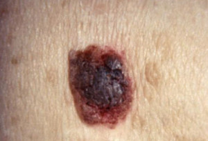
Melanoma. Shutterstock/Nasekomoe
“Blue/white veil [under magnification], peripheral globules, expansion of pigment network, inverse pigment network, etc., have all been studied and shown to be worrisome for melanoma, but there are melanomas that have no pigment.
“There are multiple types of melanoma that grow in different patterns as well, and often can look like thick, vascular lesions or seborrheic keratoses.
“SKs often are thicker with no distinct pigment network and crypts. Again however, these lesions can be flat, have an apparent pigment network and become inflamed.”

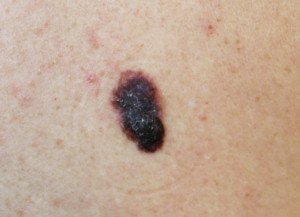
Seborrheic Keratosis
How easy can a doctor mistake a melanoma for a seborrheic keratosis after viewing it through a handheld lens (dermatoscope)?
Dr. Gordon explains, “Dermatoscopes polarize light and magnify, making it easier for us to see the intricacies of lesions, but it does not have a ‘melanoma’ light that pops up when we see one.
“All it does is give us more information about what the lesion looks like, and as we gain more research about different features that distinguish malignant lesions from benign lesions, we can continue to use these to help us decide whether to biopsy.
“However, a good rule of thumb is that the dermatoscope should never talk you out of taking a biopsy, but if you see something abnormal that you could not see with the naked eye, it is then useful to help push you towards taking a biopsy.
“Again, although there are a few features that are either reassuring or worrisome, many lesions contain both types of features.”
Best way, short of biopsy, to indicate possibility of seborrheic keratosis vs. melanoma?
“Another very important thing to remember is that we are seeing a snapshot of your skin lesions,” says Dr. Gordon.
“Change and evolution of lesions is an extremely important factor in malignancy, so monitoring and watching for change is another important job for both you and your dermatologist.”
A seborrheic keratosis can change or evolve, just like melanoma does. However, an SK’s changes can be rapid, noticeable by the naked eye from day to day. Melanoma changes much more slowly.
Thus, that weird “mole” that looks different on Thursday than it did Monday is very unlikely to be a melanoma—though by sheer coincidence, a melanoma can start growing within a seborrheic keratosis!
But this is pure coincidence; a seborrheic keratosis cannot cause cancer or turn into cancer.
Should a patient insist on a biopsy simply by hearing, “It’s a seborrheic keratosis”?
“We see hundreds of skin lesions a day and use patterns to help decide what we believe is worrisome or not,” says Dr. Gordon.
“This is our job and most of us are good at it. However, to Err is Human and there will be mistakes.
“If a patient is truly worried about a lesion, then biopsy it! If they think it is changing, biopsy it!
“I don’t condone sampling every pigmented lesion but if there is an ugly duckling or one a patient has been worried about, then let both of you sleep that night by putting it in a jar.”
So is the image above a melanoma or a seborrheic keratosis? It is a seborrheic keratosis — but note how similar it looks to the image of the melanoma!


