
If you suffer from mole anxiety and fear of melanoma, you need to read this helpful information for the “molechondriac.”
- Do you find yourself constantly checking your moles for changes? Imagining changes?
- Worrying incessantly about melanoma?
You should make an appointment with a dermatologist and request removal and biopsy of any moles that worry you. It’s about trusting your gut.
“Melanoma skin cancer is the most dangerous type of skin cancer and can appear as a dark, irregularly shaped mole or a dark patch of skin,” says Alberto de la Fuente Garcia, MD, a board certified dermatologist at VIDA Wellness and Beauty with 15+ years of experience.
“It may also look like a normal mole but change in shape, size or color over time.
“It’s important to be aware of any moles that change, or that look different from the rest, as this could be a sign of melanoma.”
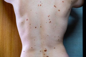
What if you have hundreds of moles? Obviously, having every one removed is not practical.
Mole Mapping Technology
A superb way to slaughter a lot of that anxiety over melanoma is to get set up with serial digital dermoscopy.

Serial digital dermoscopy
The availability of this groundbreaking technology is becoming increasingly more common.
Chances are, there’s a mole mapping clinic within reasonable driving distance of your home.
Moles are photographed; the images are input into a computer that then compares the images to a database. The moles are rated as being benign or suspicious looking.
Sometimes, though, the only comparisons that are done are by your dermatologist, who is trained at viewing amplified images of moles on a computer screen for side-by-side comparisons.
This is much more accurate than simply looking at them on your body, even with a handheld magnifying lens.
The side-by-side comparison of the same mole, taken a year to two years apart (depending on the doctor’s recommendation), can reveal so much more detail.
The computer and the human eye do NOT diagnose melanoma; it can only be suspected.
Only a pathologist can make the diagnosis after viewing cells under a microscope.
You’ll probably have to pay out of pocket. I say “probably” because people with, for instance, hundreds of dysplastic nevi (which are at increased risk for melanoma), may get an order from their dermatologist for serial digital dermoscopy — which your insurance may pick up.
Serial digital dermoscopy is especially beneficial for moles on your back that you can’t inspect closely or for people with visual impairment.
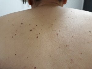
Many atypical moles. Shutterstock/Mikel Ugarte Gil
Home Mole Mapping
If you have illustration skills, you can draw sections of your body on a sketch pad, then insert your moles.
This will enable you to keep track of any new moles. You’ll no longer wonder, “Has that mole always been there?”
I suggest also drawing some of your non-mole lesions for easier reference to nearby moles.
For example, if you have angiomas (bright red but harmless spots), indicate those with a red pen.
Any moles nearby these benign spots will be easier to monitor.
These days, though, there are a number of apps that can take images of your moles and flag for suspicious signs.
However, apps should never be a replacement for an actual physician examining your body. Think of apps as an adjunct to your skin health exams.
Naming Moles Based on Their Appearance
I have a mole that’s shaped like a barn. If it begins changing, I’ll know it quickly enough, because “barn” is etched in my mind.
I have another mole that’s shaped like a bat in flight.
I have several moles that I call “two-toned” or “three-toned,” because they’re comprised of two or three shades of brown.
I have a few “diamonds,” referring to their shape.
Ask Your Dermatologist to Point out the Seborrheic Keratoses on Your Body
SK’s are benign skin barnacles. However, they may look like moles.
Ask your doctor to show you where you have these, if any.
Once you know which spots are SK’s, you won’t have to keep checking them every month. SK’s can change in appearance, but this is a benign occurrence.
Seborrheic keratoses. Shutterstock/NICKY1841
Twice a Year Skin Exams
You’ve probably read many times that you should have a dermatologist examine your skin once a year if you have an average or typical risk for melanoma.
To reduce anxiety, consider having a clinical exam twice a year.
Taking these measures will reduce a lot of anxiety and increase the chances of catching melanoma very early.
Another point to consider is that a clinical exam can reveal signs that are suspicious for other types of skin cancer.
“Skin cancer can have many different appearances, depending on the type of skin cancer and how advanced it is,” says Dr. de la Fuente Garcia.
“Non-melanoma skin cancers (such as basal cell carcinomas and squamous cell carcinomas) often appear as raised bumps that are pink or red in color.
“They may be fleshy, scaly or waxy in texture and can bleed easily when scratched.”
And by the way, before your dermatologist begins the skin exam, request that they use a dermatoscope.
 Dr. de la Fuente Garcia